Combat Arts Elbow Injuries Overview
The elbow joint is comprised of the upper arm bone or humerus and the two forearm bones called the radius and ulna. It is a compound joint: a hinge that bends and straightens the arm, combined with a pivot at the radius that lets the forearm rotate, so the palm can turn up and down.
Elbow injuries are common in martial arts as the elbow is used as a weapon, a lever for armbars, and is essential for holds and throws. This makes the elbow very vulnerable to injuries such as dislocation of bones, damaged ligaments, bursitis, bone bruises, fractures, and broken bones.
Causes of Elbow Injury
The elbow is nothing but an articulation between the humerus, ulna, and radius. This unique combination makes it both a hinge and a pivot joint. It is responsible not just for flexion and extension but pronation and supination as well. It is generally stable due to the many ligaments supporting the structure.
In MMA like BJJ, one of the first moves to learn is the armbar. In an armbar move, if held for too long or if the fighter refuses to tap out then the collateral ligaments get stretched and damaged. If the force is too much, it can tear. Over time, the repetitive stretching can make the ligaments lax and injure the joint. Falling on an outstretched arm or using the elbow strike can also damage the elbow joint.
Elbow Injury Symptoms
A badly injured elbow, particularly a dislocation, usually announces itself. The list below describes that acute picture — a dislocation or a fracture. It does not describe the slower problems this page also covers, such as tennis elbow, bursitis and tendonitis, which build over weeks and can look like nothing at all. Either way the diagnosis is made by a doctor with an x-ray, not from the side of the mat. Signs to act on include:
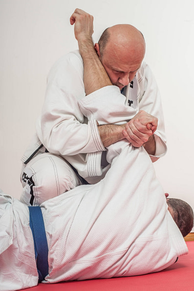
Source: guruXOX, Standard License, via Shutterstock
Types of Elbow Injuries
The elbow takes a beating in combat sports and is one of the more commonly injured joints in martial artists. Two broad patterns account for most of it. The first is an acute injury such as an elbow dislocation. The second is chronic overuse: tennis elbow, which affects the tendons on the outside of the elbow, and golfer’s elbow, which affects the tendons on the inside. They are two separate conditions on opposite sides of the same joint.
Related Injuries
Elbow dislocations typically require an extremely forceful strike or fall. They get described along two separate axes, which is easy to muddle. A dislocation is simple when no bone is broken, and complex when it comes with a fracture. Separately, it is complete when the joint surfaces are fully separated, and a subluxation when they are only partly separated. So an elbow can be simple and complete, or complex and complete, and so on.
In a partial dislocation, or subluxation, the joint surfaces are only partly separated. The deformity can be subtle, or gone by the time anyone looks, because the joint may have relocated on its own — but the elbow still will not work properly, and it still needs an x-ray, since a subluxation can come with a fracture just as a full dislocation can.
In severe dislocations, the blood vessels and nerves traveling to the lower arm and hand may be damaged.
Symptoms
Causes
An elbow dislocation is an acute event. A forceful strike may cause it — a direct kick to the elbow joint, landing wrong from a throw, or landing badly on the elbow, wrist or hand. An armbar taken past the joint’s limit, or a forceful turning motion, levers the elbow out of place. Repetitive load does not dislocate an elbow; that is the mechanism behind tennis elbow, covered below.
To learn how Elbow dislocation is diagnosed, Click here.
Tennis elbow is an overuse injury of the common extensor tendons on the outside of the elbow — lateral epicondylitis — driven by overload of the extensor carpi radialis brevis (ECRB) tendon. Golfer’s elbow is its mirror image on the inside of the elbow, affecting the common flexor tendons; everything that follows in this section describes tennis elbow. In MMA, loaded gripping with the wrist held in extension is what causes it. The elbow takes a battering in almost every attack and defense in martial arts like jiu-jitsu, and that strains the lateral epicondyle. Training with weapons also loads the elbow while the wrist extends. It starts as gentle pain and inflammation, and with time and repetitive stress it worsens.
Every time a fighter grapples, uses his elbow to do a chokehold, or defends, the elbow joint is stressed. Degenerative changes take place in the ligaments. Microtears occur in the fibers which causes inflammation. The repair tends to be disorganized and hasty given that the joint experiences continual trauma. The repeated wrist extension, radial deviation, and forearm supination gradually worsen the condition.
To learn how Tennis elbow are diagnosed, Click here.
Golfer’s elbow — medial epicondylitis — is an overuse injury of the flexor-pronator tendons where they attach to the bony bump on the inside of the elbow, the medial epicondyle. It is the mirror image of tennis elbow and is less common. In martial arts it is linked to repeated, forceful gripping with the wrist flexed: gi grips on the collar and sleeve, frames and posts, and weapons work.
Pain and tenderness on the inside of the elbow that can spread down the inner forearm, pain when gripping, making a fist or bending the wrist against resistance, and a weaker grip. Numbness or tingling in the ring and little fingers suggests the ulnar nerve, which runs just behind the medial epicondyle, is involved too and should be mentioned to a doctor.
Repeated gripping with the wrist flexing and the forearm turning palm-down loads the flexor-pronator tendons faster than they can adapt. Sudden increases in grappling volume, heavy grip training and too little recovery between sessions add to it. As with tennis elbow, the change in the tendon is mainly degenerative rather than inflammatory.
To learn how Golfer’s elbow is diagnosed, Click here.
Common Elbow Injuries
Other common injuries involving the elbow include tendonitis and bursitis. Read about them in the Common Injuries section.
Elbow Injury Diagnosis
Elbow dislocations are usually diagnosed with radiography. This is to rule out any fractures involving the humerus, radius, and ulna. A physical exam is important to understand the limitations of the joint. Tennis elbow is more often a clinical diagnosis.
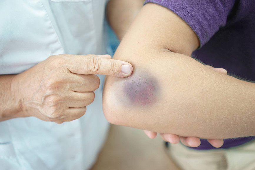
Source: Nadezhda Novikova, Standard License, via Shutterstock
Injury Specific Diagnosis
Physical Exam
The first step in the physical exam is to visually inspect the joint and look for a visible deformity. If the joint is malpositioned, then an extensive exam is required.
The skin is inspected for ecchymosis and the joint is palpated for effusion. The elbow is then palpated for tenderness. The wrist is palpated for instability. This is to rule out any involvement of the radioulnar joint. Articulations of the joint are examined via a cautious range of motion tests. Although neurovascular injuries are rare, it is important to examine the nerves and pulses.
To this end, the motor nerves are tested individually. The patient is asked to make a fist, to tests the flexor digitorum superficialis. The doctor may ask the fighter to place the fingers flat on the table and then flex a finger. To test the flexor pollicis longus, the fighter is asked to make an “OK” sign. The doctor simultaneously evaluates the flexion of the thumb at the interphalangeal joint. The radial nerve is evaluated by asking the fighter to extend their wrist. If unable to so, it is also called a wrist drop. The fighter is asked to make a “thumbs up” gesture to test the posterior interosseus nerve. For the ulnar nerve, the fighter is asked to abduct and adduct their fingers.
Imaging
Doctors will want to understand the type of dislocation before they treat it. X-rays are done. These include both anteroposterior and lateral views. To identify the direction of the dislocation and rule out any surrounding fractures oblique views of the elbow are helpful. After the reduction, Xrays are repeated. CT is done only if there are associated injuries.
Lab Tests
Lab tests are usually unnecessary, since most elbow dislocations are reduced without an operation. If surgery is warranted, then pre-operative blood tests are required.
To learn how a Elbow dislocation is treated, Click here.
Physical Exam
Since this is a chronic condition, fighters usually complain of pain for a long time. A detailed history of use and overuse is reviewed. The lateral elbow is palpated for the point of maximal tenderness. The usual site of tenderness tends to be a focal, distal location about 1-2 cm from the lateral epicondyle.
On wrist extension, if the pain is reproduced or increased then it’s indicative of tennis elbow. This is more diagnostic if the elbow is extended and the forearm is pronated. On exam, if while resisting the extension of the middle finger, with elbow extension, there is pain, the diagnosis is confirmed.
Imaging
This is a clinical diagnosis. Usually, imaging is not necessary. An elbow radiographic series with anteroposterior and lateral views are ordered to rule out other injuries. Ultrasound and MRI are used to identify degenerative joint changes, fractures, bursitis, tears, stress fractures, or osteochondral defects.
Lab Tests
Lab tests are not part of diagnosing tennis elbow. Despite the "-itis" in the name, the change in the tendon is degenerative rather than inflammatory, and inflammatory markers such as ESR and CRP are not routinely ordered for it — they are used when a doctor suspects something else, such as an inflammatory arthritis or an infection. If surgery is warranted, then pre-operative blood tests are required.
To learn how Tennis elbow are treated, Click here.
Physical Exam
Doctors press over and just below the medial epicondyle to find the point of greatest tenderness and check whether the pain is reproduced by bending the wrist or turning the forearm palm-down against resistance. They also examine the ulnar nerve and test the ulnar collateral ligament, because both can cause pain on the inside of the elbow.
Imaging
Golfer’s elbow is a clinical diagnosis and imaging is often not needed. X-rays may be taken to rule out other problems, and ultrasound or MRI can confirm tendon changes or look for a tear, a ligament injury or another cause when symptoms do not settle.
Lab Tests
Lab tests are not part of diagnosing golfer’s elbow. They are only used when a doctor suspects something else, such as an inflammatory arthritis or an infection.
To learn how Golfer’s elbow is treated, Click here.
Diagnoses of Common Elbow Injuries
To diagnose elbow injuries a variety of imaging modalities are helpful. Read more about them in our Common Diagnoses section.
Elbow Injury Treatment
Treatment depends on the type of injury and severity. A dislocated elbow is put back by a doctor, and x-rays come first wherever that is practical: an associated fracture or a trapped fragment changes what should be done, and forcing a reduction on a fractured elbow can do lasting harm. Nobody at ringside or in the gym — coach, corner, training partner or the fighter — should attempt to put an elbow back in. Get to an emergency department. Chronic injuries require the fighter to stop the offending activity, and surgery is reserved for cases where symptoms do not improve and conservative treatment fails.
Injury Specific Treatment
Emergency Treatment
An elbow dislocation is an emergency. Check for a pulse at the wrist and for normal sensation in the hand, because the displaced bones can compress or tear the vessels and nerves running to the forearm and hand. An absent pulse, a cold or pale hand, or numbness means go now rather than later. Support the arm in the position it is already in, keep the fighter still, and get to an emergency department. Do not pull on it, and do not try to force it back.
Medical Treatment
The first step is to attempt a closed reduction of the elbow. A reduction will decrease the pain and swelling of the joint. Often, reduction requires sedation. If the elbow is displaced, then the displacement is first taken care of before applying longitudinal traction to the forearm.
Since posterolateral dislocations are more common, the elbow is extended. The doctor applies slight pressure on the posterior side of the olecranon. This reduces the elbow. A palpable or audible clunk is heard during reduction. After the elbow is reduced, all range of motion is tested which involves, flexing, extending, valgus, varus, pronating, and supinating. Then the elbow is placed in a posterior long-arm splinting. This is 90 degrees of flexion.
Two weeks later, the splint is removed. Physical therapy is then started. For less stable joints, the splint will be kept for longer with the help of extension blocking splints. Avoid prolonged immobilization.
Surgical management is rarely required. If a doctor cannot reduce the dislocation, then closed reduction under anesthesia is advised. If the elbow is unstable at 50 to 60 degrees of extension, then surgery is warranted. The goal is to allow a range of motion of 30 to 130 degrees in flexion and extension and 50 degrees of pronation and supination. If there are unstable fractures with a complex dislocation, surgery is necessary. This could be due to fragments within the joint or entrapped medial epicondyle in the joint.
Home Treatment
After reduction, splinting is done with a posterior long arm splint. This prevents the extension of the elbow. Elbow splinting is usually in 90 degrees of flexion. For pain, analgesics and NSAIDS are helpful. Physical therapy is started after two weeks if the joint is stable.
Emergency Treatment
Tennis elbow is generally not an emergency. The pain and symptoms usually develop over time. An acute injury is precipitated due to chronic strain. In case of an acute injury, rest, ice, compression, and elevation are essential followed by orthopedic consult.
Medical Treatment
The first step is nonoperative management for tennis elbow. This includes removing the offending movement that causes pain. To achieve pain control ice the area after any physical activity. Use topical NSAIDS for escalating pain. Counterforce straps like "Cho-pat" straps relieve tension at the lateral epicondyle. Wear these forearm straps while doing all activities. A brace is also useful. Braces like a cock-up wrist splint can ease the tension off the wrist extensors.
Surgical Management
Surgery is usually the last resort in tennis elbow. Non-operative management is prescribed for a period of 6- 12 months. If it fails, then surgery is warranted. ECRB debridement and release of the tendon origin at the lateral epicondyle is a popular technique. To stimulate healing potential, the pathologic tissue must be debrided leaving behind bleeding, a bed of bony tissue.
Home Treatment
In tennis elbow, therapy plays an especially important role — both occupational and physical therapy. The focus is usually on stretching the forearm and strengthening the muscles, with gradual progression to eccentric loading of the common extensor tendon. If the pain is not settled by any of that, some clinicians offer injection or patch treatments: platelet-rich plasma, botulinum toxin, prolotherapy, or nitrate patches. The evidence for all of them in tennis elbow is limited. Botulinum toxin in particular works by temporarily weakening the wrist and finger extensors, which for a fighter means a spell of reduced grip and wrist strength. Weigh that trade-off with an orthopedic surgeon before agreeing to any of them.
Emergency Treatment
Golfer’s elbow is not an emergency. Get the elbow checked promptly if the pain started with a sudden pop during a throw or armbar, if the elbow feels unstable, or if numbness or weakness in the hand is getting worse.
Medical Treatment
Treatment starts with cutting back the grips and loads that provoke the pain rather than stopping training altogether, with ice or simple pain relief for flare-ups and, for some people, a counterforce strap. Physical therapy with progressive strengthening of the wrist flexors and forearm is the mainstay. A corticosteroid injection can ease pain in the short term, but the benefit tends not to last and repeated injections are not advised. Surgery is reserved for the few whose symptoms persist after 6 to 12 months of well-managed non-surgical care.
Home Treatment
Most cases settle over several months. Modify grip-heavy drills, rebuild grip and forearm strength gradually, and increase grappling volume in steps. Mild discomfort during rehab is acceptable if it settles by the next day; pain that lingers means the load went up too fast.
Common Treatments
Elbow injuries are treated conservatively and surgically. Some common medications and therapies are listed in our Common Treatments section.