Combat Arts Foot Injuries Overview
The human foot provides the body with structural strength, a foundation for movement, shock absorption, and locomotive power. Each foot holds around 26 bones and roughly 33 joints, bound together by about a hundred ligaments, tendons and muscles — which is why the two feet between them account for roughly a quarter of all the bones in the body. Medically speaking, the ankle is part of the foot, although most people refer to them as unique parts.
Components of Foot
Hindfoot has three joints that link the foot through the ankle or talus. The talus creates a hinge that allows the foot upward and downward motion by connecting to the tibia and fibula. The calcaneus, or heel bone, is the largest bone in the foot; it forms the back of the foot and the bottom of the heel, where it is protected and cushioned by a pad of fat.
Midfoot forms the arch of the foot, absorbing shock and pounding during movement. It is made up of five tarsal bones, joined to the hindfoot and the forefoot by the tarsal and tarsometatarsal ligaments, and braced underneath by the plantar fascia — a thick sheet of connective tissue, properly an aponeurosis, that spans the arch.
Forefoot includes the five metatarsals and the toe bones, or phalanges, which together provide flexibility, strength, and movement. The forefoot helps keep the pressure on the ball of the foot evenly balanced, and it takes a large share of the load every time you push off. The toes are particularly prone to injury because of their many small bones and their primary position in movement propulsion and supporting the body's weight.
Former Strikeforce Heavyweight Champion Alistair “The Demolition Man” Overeem has held world titles in both MMA and K-1 kickboxing, and he has also been sidelined by an injury to a toe — a reminder that the smallest bones in the forefoot can stop a heavyweight. Toe injuries have their own section on this site and are not covered on this page.
Foot Injury Causes
Issues with the feet may result in problems elsewhere in the body, such as pain in the hips, back, or legs. Conversely, problems, or weaknesses in other parts of the body will often affect the feet. For instance, when hip abductors, psoas, or back muscles are weak or injured, often the feet will respond to these issues with pain, gait problems, bunions, hammertoe, or other difficulties.
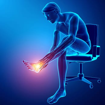
Foot pain may occur because of structural issues in joints, ligaments, tendons, fascia, muscles, or bones, be referred pain from a pinched nerve in the back, neck, or hip, or may occur because of systemic diseases such as diabetes. The diagnoses behind that pain include stress fracture, toe injury, plantar fasciitis, bunions, heel spurs and Achilles tendinosis. Forefoot or midfoot pain that builds over weeks of training, hurts when you press on one spot of bone, and eases with rest deserves particular attention — navicular and metatarsal stress fractures are easy to mistake for metatarsalgia, and they are slow to heal if they are missed. It is essential to consult with a health professional or foot specialist when foot pain occurs and especially when it is ongoing.
Foot Injury Symptoms

Source: Marko Aliaksandr, Standard License, via Shutterstock
Types of Foot Injuries
Since the foot is composed of many bones, and a host of ligaments and tendons, any number of the structures are prone to injury. It also bears all the weight irrespective of the type and speed of motion. Some of the injuries could be due to acute injuries, others due to chronic wear and tear. This repeated wear can lead to degenerative changes in the fascia and tendons. Some of these injuries include Plantar fasciitis, bunions, metatarsalgia, and heel spurs.
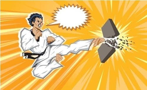
Something as Small as a Foot Injury Limit Your Combat Goals
Sign up Today to Prevent Those Troublesome Injuries!
Related Injuries
This is a repetitive strain injury that creates persistent pain at the bottom of the foot. The pain of plantar fasciitis is felt mainly at the heel, where the fascia attaches to the heel bone, and it can radiate forward into the arch. The foot has a thick, wide fibrous band of connective tissue called fascia that stretches from under the heel to the front of the foot, spanning the arch of the foot, and helping to support the arch. This is the plantar fascia or plantar aponeurosis, and it is strong like a ligament, connecting bones at the heel of the foot to bones at the front of the foot.
These tissues are made up of three segments originating at the base of the heel bone. The middle section provides shock absorption to the foot while running and base support when standing. Martial artists may develop pain in all three sections, but typically, it is this center fascia that becomes most tender and painful.
Causes
The causes of plantar fasciitis, or plantar fasciosis, are not completely understood, but they appear to involve overuse and repetitive overloading of the fascia, producing small tears and, over time, degeneration of the collagen where the fascia attaches to the heel bone. It was originally thought to come from inflammation in the tissue and was named plantar fasciitis, since “itis” means inflammation.
The pain is from deterioration of the tissue due to overuse, now referred to as plantar fasciosis. In 2003, Lemont et al looked at 50 cases of plantar fasciitis and found so little evidence of inflammation that they declared plantar fasciitis “is a degenerative fasciosis without inflammation, not a fasciitis.”
Symptoms
The primary symptom of plantar fasciitis is heel pain, felt under the heel and often spreading forward into the arch. The pain usually feels like a sharp stabbing, and it is typically most intense when first getting up in the morning or after sitting for long periods.
To learn how Plantar Fasciitis is diagnosed Click Here
Plantar fasciitis and heel spurs are usually discussed together, and the pain is not a reliable way to tell them apart — both are felt at the heel. A heel spur is bone that has formed where the fascia or the Achilles tendon attaches to the heel, and spurs turn up on imaging in plenty of people who have no heel pain at all. The two conditions are associated, but a spur has not been shown to cause plantar fasciitis, or the other way round.
Heel spurs are small outgrowths of bone at the attachment of the plantar fascia or Achilles tendon on the heel — bone laid down within the attachment, not a deposit of calcium sitting on it. They are associated with repetitive stress from pounding, walking, running and kicking, and with sustained pull from injured tendons and ligaments.
Symptoms
Pain and sensitivity to touch are the most common symptoms, but some people may be asymptomatic. The pain comes from irritation of the soft tissue around the calcification area. The pain may feel achy or like a knife or pin sticking into the bottoms of the feet. Often the pain eases after movement and is typically due to inflammation.
Causes
Martial artists who run, jump or kick repeatedly may cause heel trauma which incites the body’s healing mechanism to produce bone or heel spurs. Heel spurs usually occur when there is already chronic pain experienced through the heel or foot arch because of chronic inflammation, thickening, and microtears of the connective fascia tissue resulting in plantar fasciitis.
To learn how Heel Spurs is diagnosed Click Here
This is a general term meaning pain in the ball of the foot. The metatarsals are the five long bones that run forward from the midfoot to the base of the toes; it is the talus and the heel bone, not the metatarsals, that connect the foot to the ankle. Metatarsalgia is mechanical overload pain at the metatarsal heads — the ball of the foot taking more punishment than it can absorb, as from repeatedly kicking boards or a heavy bag. That is a different problem from a Morton's neuroma, where a nerve between the metatarsals becomes thickened and irritated, and the pain is more burning or electric, often with numbness in two neighbouring toes. The body weight is balanced through this area by many small bones that may cause simple discomfort up to extreme pain when out of balance or injured.
Typically, the pain will be worse while on the feet and moving. It may hurt to flex the toes and feel pain when wearing shoes, socks, or other gear that surrounds and applies pressure on the foot. Pain may be felt in the ball of the foot when running, jumping, kicking, and other physical activities.
The primary symptoms typically felt in the ball area of the foot include:
Metatarsalgia, or pain in the ball of the foot, may come on while throwing a much heavier opponent or driving a powerful BJJ takedown, or from high tornado kicks landed badly on the ball of the foot, bruising bone and overloading the metatarsal heads. Any overuse, trauma, or injury to the foot may cause metatarsalgia. Examples include:
To learn how Metatarsalgia is diagnosed Click Here
A bunion is a deformity or hard bony bump that forms typically at the base of the first MTP joint or big toe on the inner edge of the foot, although bunions may also develop on the fifth MTP joint or little toe MTP joint on the outside of the foot. The MTP or metatarsophalangeal joints are located at the base of the toe and connect the first metatarsal toe bones to the foot bones. These MTP joints help hold and distribute weight when the body is standing or moving. MTP joints have a thin joint capsule which surrounds them, and ligaments which connect and support them. The first MTP joint or big toe joint is the most prone to developing bunions.
A bunion develops when the first metatarsal bone of the foot starts to shift its position from straight forward to angle off to the side. The big toe then leans into and presses against the other toes which cause an unnatural and painful angling of the joint. A bunion usually feels painful from the beginning and becomes progressively more so as the protruding bone increases in angle and size due to swelling, restricting the movement of the toe, foot, and body.
As a bunion increase in size, it also increases pressure upon other toes and may inflict damage at this primary juncture. Pressure on bones, ligaments, and tendons in this area can impair the foot's functioning and create a condition called hammer toe, or the bursa, which are cushioning fluid-filled sacs or pillows between joints may become sore, inflamed, or infected.
Martial artists, whose feet are constantly stressed from kicking, standing, running, jumping, or moving on the feet for long periods are susceptible to developing bunions. They are also likely to develop bunions because of physical stress placed on feet from repetitive movement, pounding, and often badly fitting shoes. Most shoes don't allow sufficient room for a bunion and place added pressure on the swollen and misaligned joint, increasing pain and promoting further deformation of the area.
Bunions also run in families because certain foot shapes, for example, narrow or wide feet, foot structures such as high or low arch, the shape of the metatarsal head on the toe joint, long or short toes are all hereditary, and some types develop bunions more easily than others. Feet that have loose joints and tendons, long toes, flat feet, low arch, and certain other characteristics are more likely to develop bunions. Bunions may develop from obesity, as well, since excess weight places pressure on the toe joints.
To learn how Bunions are diagnosed Click Here
A stress fracture is a small crack in a bone caused by repeated loading rather than a single blow. In the foot, the second and third metatarsals are the most common sites, followed by the navicular and the fifth metatarsal. It starts as a stress reaction in the bone and can progress to a complete fracture if training carries on. Navicular and fifth metatarsal stress fractures are treated as high-risk because they heal poorly and are more likely to need a period off weight-bearing or surgery.
Symptoms
Pain that builds over days or weeks of training. At first it comes on during activity and eases with rest; later it starts earlier in each session and may ache at rest or at night. There is usually one spot on the bone that hurts to press, sometimes with mild swelling on the top of the foot. A navicular stress fracture often causes a vaguer ache on the top or inner side of the midfoot that is easy to dismiss.
Causes
Stress fractures happen when bone is loaded faster than it can adapt: a sudden jump in training volume or intensity, extra roadwork during fight camp, repeated jumping and kicking on hard floors, or a change of footwear or training surface. Not eating enough for the training load, which is common during weight cuts, low vitamin D, missed periods, and a previous stress fracture all raise the risk.
To learn how Stress Fractures are diagnosed Click Here

Source: Liderina, Standard License, via Shutterstock
Common Injuries
In our Common Injuries section, we discuss common injuries such as foot sprains and contusions.
Foot Injury Diagnosis
Various tests and special exams are needed to diagnose foot injuries. Imaging supplements the physical exam and confirms the diagnosis.
Injury Specific Diagnosis
Physical Exam
Doctors will try to reproduce the pain by palpating the plantar medial calcaneal tubercle. They will also try to reproduce the pain with passive dorsiflexion of the foot and toes. This passive dorsiflexion of the first MTP joint is known as the windlass (or Jack's) test.
It is considered a positive test if there’s pain. The doctors will examine gait and look for fat pad contusions, fat pad atrophy, stress fractures, and nerve entrapments.
Imaging
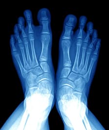
Plantar fasciitis is a clinical diagnosis. Imaging is not required. Yet, plain films are done to evaluate other injuries. This is especially if the injury persists. X-rays and ultrasound evaluations show calcifications in the soft tissues or heel spurs.
An ultrasound will also show thickening and swelling of the plantar fascia. MRI is only requested if conservative therapy fails. It can visualize tears, stress fractures, or osteochondral defects.
Lab Tests
Blood tests are not required to diagnose plantar fasciitis.
To learn how Plantar Fasciitis is treated Click Here
Physical Exam
Doctors will view the foot first and check for any swelling, atrophy, lack of sensation, and skin tears. They will examine the range of motion of the ankle and foot. They will check the gait, arches of the foot, atrophy of the heel pad, and foot alignment. They will palpate the plantar fascia, spurs, tenderness to them, and aggravation by bearing weight. Muscle strength is tested and exams specific to the calcaneus are done. Once such an exam is the calcaneus squeeze test. This is a test where a compressive force is applied to the calcaneus. If pain is reproduced, then the test is positive.
Imaging
Calcaneal spurs are diagnosed with plain films, CT, and MRI. They appear as bony projections that are seen either at the origin of the plantar fascia or the insertion of the Achilles tendon.
Lab Tests
Blood tests are not required to diagnose calcaneal spurs.
To learn how Heel Spurs is treated Click Here
The doctor will evaluate the plantar foot and evaluate the callus areas where there is an increase in foot pressure. They will check the arch of the foot. The metatarsals are evaluated during the gait cycle as well. The calf muscle is examined for tightness.
The diagnosis is confirmed by combining physical examination and imaging. The imaging includes ultrasound and MRI. MRI can provide a detailed image of soft tissues and any early osteophyte changes.
Lab tests are not necessary to diagnose metatarsalgia. The physical exam combined with imaging is necessary to diagnose it.
To learn how Metatarsalgia is treated Click Here
In the physical exam, doctors will look for the deformity and evaluate whether there is subtalar joint stiffness, midtarsal joint stiffness, a particular resting calcaneal stance position, and tibial torsion. They will also look for any forefoot varus. They have to identify whether the deformity is present only on weightbearing. They will assess the position of the hallux compared with the second toe, and look for any lateral deviation of the MTP joint.
The ROM of the first MTP joint range of motion is assessed. The quality of the joint is assessed whether it has pain or not. Then its axis of motion is checked. The first MTP undergoes two assessments; resting position and range of motion and the second is mobility.
Weightbearing deformities are visible. They are accompanied by increased hallux abduction, increased medial prominence, MTP joint dorsiflexion, hallux purchase, and metatarsus varus.
The diagnosis is usually made by a physical exam. Imaging will identify the extent of joint damage. The imaging is done by plain films with X-rays (AP and lateral weight-bearing) of the foot. Imaging displays lateral hallux deviation at the first MTP. This guides the treatment procedure.
The classification of the valgus is mild, moderate, and severe. It is based on the films obtained through the weight-bearing anteroposterior, lateral oblique, lateral, and sesamoid axial views. These views also help view the structural status of the foot.
The AP projection assesses all the angles, the hallux rotation, and the condition of the first MTP joint.
The lateral projection assesses the position of the joint and dorsal osteophytes. The lateral oblique projection evaluates the trabeculation and projection of the bone. The sesamoid axial view assesses the degenerative joint changes.
Imaging provides the Degree of the Hallux valgus angle (HVA) / Intermetatarsal angle (IMA)
Normal: <15⁰ / 9⁰
Mild: 15-30⁰ / 9-13⁰
Moderate: 30-40⁰ / 13-20⁰
Severe: >40⁰ / > 20⁰
If doctors suspect osteomyelitis, MRI, and radionuclide imaging is done. However, this is very rare.
Lab Tests
Lab tests are not required for HV deformities. However, if metabolic conditions are suspected then Rheumatoid factor, ANA, CRP, ESR, uric acid, and CBC.
To learn how Bunions are treated Click Here
Physical Exam
Doctors ask about recent changes in training, diet and weight, and about previous stress fractures. They press along the metatarsals and over the navicular to find a point of bony tenderness, look for swelling, and may ask the fighter to hop on the affected foot, which often reproduces the pain.
Imaging
X-rays are usually taken first, but they are often normal for the first two to three weeks, so a normal x-ray does not rule a stress fracture out. MRI is the most sensitive test for an early stress injury and shows how severe it is. CT is used to look closely at navicular and other high-risk fractures, and a bone scan is an alternative when MRI is not available.
Lab Tests
Blood tests do not diagnose the fracture itself. After repeated stress fractures, or when under-fuelling is suspected, doctors may check vitamin D, calcium and hormone levels and may recommend a bone density scan.
To learn how Stress Fractures are treated Click Here
Source: Shutterstock, Standard License
Common Diagnoses
Learn more about how foot injuries are diagnosed and confirmed in our Common Diagnoses section.
Foot Injury Treatment
There are various treatments used to treat injuries in the foot. A vast array of therapies to treat the fascia, the bones, the ligaments, and tendons. The cornerstone of every therapy is a good physical therapy program which includes stretching, condition, and strengthening.
Injury Specific Treatment
Emergency
Plantar fasciitis is not a medical emergency. The pain is excruciating enough to make it feel like it is. The first step is managing the pain.
Medical
The offending activity must stop. Relative rest is advised. Ice and topical NSAIDs are used for pain management.
Other alternatives like deep friction massage of the arch and insertion are beneficial. Shoe inserts or orthotics and night splints are also recommended. Stretching and rehabilitation of the plantar fascia, Achilles' tendon, gastrocnemius, and soleus are emphasized in every treatment protocol.
If the pain is not relieved by conservative treatment, then extracorporeal shock-wave therapy, botulinum toxin A, autologous platelet-rich plasma therapy, dex prolotherapy, and steroid injections are other options. Surgery is only advised in chronic conditions and if other therapies have failed. Every therapy though will need at least 6 weeks to have any effect.
Alongside this, stretching exercises, ice packing, and heel strapping are used to settle pain and unload the fascia — bearing in mind that the underlying problem is usually degeneration of the tissue rather than true inflammation, which is why loading the tissue progressively matters more than chasing swelling. A night splint is also helpful provided the offending activity is halted.
Surgical options include fasciotomy via endoscopy or it could also be via an open approach.
Home Treatment
Plantar fasciitis usually settles over months rather than weeks, and most people get better without surgery. That is not a reason to do nothing: recovery tends to be quicker and relapse less likely with rest from the aggravating activity, arch support from good shoes or inserts, and a progressive stretching and loading programme — which is why the medical section above calls a physical therapy programme essential. Most martial artists with this condition should consider wearing a night splint. Heel pain that changes character deserves a fresh look rather than more of the same: pain at night or at rest, numbness or pins and needles, or pain that keeps building despite rest can mean a calcaneal stress fracture or a nerve entrapment rather than plantar fasciitis.
Fighters must learn to stretch the plantar fascia with basic home exercises. Wear appropriate shoes with adequate arch support, preferably made by a podiatrist. Avoid walking barefoot, lose weight, and curtail repetitive exercises that traumatize the heel.
Emergency Treatment
This is not a medical emergency. These calcaneal spurs develop over a long period and hence can be treated conservatively.
Medical Treatment
Conservative treatment works for most people and surgery is rarely needed. Simply resting the foot and heel for a time often results in significant improvement.
Heel pain that lasts over a month may require more extensive treatment. Treatments may include: physical therapy, stretching the area, exercise, shoe inserts or orthotic devices to correct biomechanical imbalances, specialty shoes to cushion and support the area, over-the-counter pain relievers, night splints to maintain elasticity in the area, or cortisone injections. Surgery is a last resort after a long course of conservative care has failed, and current procedures address the fascia rather than simply removing the spur, which is often not the source of the pain in the first place.
Home Treatment
Use of orthoses and shoe inserts can help, Night splints must be used and pain relief through medication is helpful.
Metatarsalgia is a chronic condition and needs conservative treatment initially.
Physical therapy, orthoses and stretches form the mainstay of treatment. Conservative management is done with NSAIDs to control pain. Special orthotics must be worn with rocker sole modification and metatarsal pads. Any abnormal subtalar eversions must be corrected. MT pads can be of rubber, polyurethane, or silicone. A forefoot cushion or an MT bar can also help. Stretches in the physical therapy program must involve the calf muscles, ankles, toes, and the Achilles tendon.
Shoes and orthoses with good arch support are helpful. Metatarsal pads also relieve pressure and redistribute it across the joint.
Most bunions do not require medical treatment. Conservative treatment includes modifying shoes so that they’re low heeled and wide. Wearing orthoses to improve support and alignment can help. Analgesics like NSAID’s can relieve pain. Ice packs can reduce inflammation. Medial bunion pads prevent irritation in the affected joint. Performing stretching exercises can restore joint mobility.
See a doctor, podiatrist or orthopedic foot specialist if pain and/or swelling persists, keeps getting worse, disrupts ordinary activities, or if all shoes cause pain, since bunions can lead on to other problems: bursitis, where the small fluid-filled cushion over the joint becomes inflamed; hammertoe, where the affected toe becomes deformed; or metatarsalgia, pain in the ball of the foot. Bursitis is inflammation, not infection — but a bursa that turns hot and red, becomes rapidly more painful, or comes with a fever may be infected, and that needs to be seen urgently rather than written off as an angry bunion.
Pain is the guide for further treatment. If the joint still hurts, surgical procedures are advised. Osteotomy is a surgical procedure where the bone is cut and realigned. A Wilson osteotomy has a straight cut. A chevron osteotomy has a wedge-shaped cut. Cuts could be made to the base, shaft, or the neck. Correction usually improves the angle of the big toe over the following year, but a normal angle is not guaranteed and recurrence over time is well documented.
Arthroplasty relieves the pain and keeps the joint mobile. Here the joint is replaced by an implant. Hemiarthroplasty is where the toe length remains the same. There is less bone resection.
Arthrodesis is where the MTP joint is fused into the correct position. It's only done when the joint has undergone severe degeneration.
Soft tissue procedures such as the modified McBride release the tight structures on the outer side of the joint and tighten those on the inner side. The original McBride also removed the fibular sesamoid; that step has largely been abandoned because it lets the big toe drift the other way into hallux varus.
Home Treatment
Conservative measures typically will improve bunions. These conservative steps include:
Emergency Treatment
A stress fracture is not usually an emergency, but stop running, jumping and kicking on the foot as soon as one is suspected and get it assessed within a few days. Training through the pain can turn a stress reaction into a complete fracture. Get urgent care if the foot looks deformed, you cannot put weight on it, or the pain came on suddenly with a snap.
Medical Treatment
Most low-risk metatarsal stress fractures heal with relative rest: a stiff-soled shoe or walking boot and several weeks away from impact, with pain-free cross-training such as swimming or cycling to keep conditioning. Navicular and fifth metatarsal stress fractures are managed more cautiously, often with a period of no weight-bearing in a cast or boot, and some need surgical fixation. The clinician should also look at why it happened, including training load, footwear and nutrition.
Home Treatment
Return to training in stages once walking is pain-free: hopping, then light running, then kicking and jumping drills, building the load over several weeks and dropping back a stage if the pain returns. Eat enough to match your training, including enough calcium and vitamin D, and avoid hard weight cuts while the bone heals.


Source: Andrey_Popov, Standard License, via Shutterstock
Common Treatments
In our Common Treatments section we discuss the various options and therapies available for the treatment of foot injuries.