Combat Arts Wrist Injuries Overview
The wrist is one of the most commonly injured areas for martial artists. Damage to any part of the wrist can cause pain and affect your ability to use the wrist and hand. Hand and wrist injuries turn up often across combat sports such as BJJ, karate, tae kwon do, and MMA, though the reported rates vary a lot between sports and between studies, so nobody can honestly rank them.
The wrist is composed of eight bones, called the carpals, connected into two rows by ligaments. This arrangement gives the wrist its suppleness, strength, and ability for fluid movement.
These bones are vulnerable to damage from too much pressure such as during a fall or wrist lock which may permanently cripple the wrist or hand. A great resource to own for any martial artist, trainer, or coach for diagnosing and treating wrist and hand injuries is Hand and Wrist Injuries in Combat Sports: A Guide to Diagnosis and Treatment, by Luchetti, Pegoli, et al.
It is not uncommon to hear that a martial artist broke their wrist. Jet Li is said to have broken his right wrist while filming The Master, with the choreography reworked around it. Stories like that get repeated with no medical source behind them, and they are a bad model to copy — training or fighting on a broken wrist risks displacing the fracture and cutting off the blood supply the bone needs to knit.
Causes of Wrist Injury
Underlying causes such as repetition, sports routines, unequal ligament, tendon, or muscle strength surrounding the joint, or weakness because of previous accidents may cause pain or damage. A wrist injury can occur when striking, falling, doing dive rolls, incorrectly performing a strike with a bent wrist, improperly performed pushups, or too many handstands.
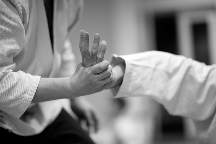
Source: Anna Jurkovska, Standard License, via Shutterstock
These types of actions can overwork and inflame the tissues around the joints or cause stress fractures, especially when performing the same repetitive movement regularly. Falling onto an outstretched hand or arm, using heavy weights improperly, or throwing incorrectly may all cause wrist pain and sprains, strains, breaks, or fractures.
Wrist Injury Symptoms
An injury to the wrist may exhibit different types of pain, depending on the cause. Pain from not warming up properly may feel like a mild tweaking or aching along one side of the wrist or hand. Repetitive use may create carpal tunnel syndrome which causes a variety of symptoms, including feeling of numbness, tingling, weakness, difficulty moving, or pins-and-needles sensations, especially at night.
Types of Wrist Injuries
The wrist takes a beating in MMA. Improper punching or fighting technique causes injuries. Grappling and excessive joint manipulation can cause sprains and fractures in the wrist bones. Years of training can also cause degenerative changes that put the wrist at further risk. The following injuries are common in the wrist: carpal tunnel syndrome, ganglion cysts, fracture of the scaphoid bone, and Kienbock's disease.
Wrist Injuries Are Very Common in Combat Arts
We Can Teach You How to Avoid These Troublesome Injuries
Related Injuries
Any MMA artist knows that for combat, the full range of motion of the wrist, fingers, and thumb is necessary. Repetitive flexion of the wrist raises the pressure inside the carpal tunnel, and that pressure squeezes the median nerve. Carpal tunnel syndrome is the most common entrapment neuropathy — it is usually put at around 90% of all entrapment neuropathies, which is not the same as 90% of all nerve disorders.
Symptoms
Causes
The carpal tunnel is a very narrow passageway that connects the wrist to the hand. Tendons, ligaments, vessels, and nerves pass through it. Carpal tunnel results from a combination of compression and traction. In MMA, various moves like grappling, throwing, grabbing, increase the pressure, obstruct venous outflow, increases local edema and compromises the median nerve's intraneural microcirculation.
So nerve function is compromised. The structural integrity of the nerve is affected by repetitive insult. The myelin sheath develops lesions over time. Gradually, the surrounding tissues become inflamed. They lose their protective and supportive ability.
The repetitive traction and wrist motion in MMA exacerbate the condition and further injures the nerve. Simultaneously, if any of the nine flexor tendons that pass through the carpal tunnel are inflamed, they can compress the median nerve.
To learn how Carpal Tunnel is diagnosed Click Here
A ganglion cyst is sometimes called a Bible cyst, from the old habit of smashing it flat with a heavy book. Do not do that. Hitting a cyst can fracture the bone underneath and damage tendons and nerves, and the cyst usually comes back anyway. A ganglion is not a tumor — it is a sac of thick mucin, and it is the most common soft tissue mass of the hand and wrist. They result from the repetitive microtrauma of martial arts that load the hands, which degenerates the connective tissue. They are also sometimes found in the knee and foot.
Symptoms
Causes
Ganglion cysts make up most of the soft tissue lumps found in the wrist, and most of them sit on the back of it. They are more common in women, but any fighter putting repetitive tension through the wrist can get one. Chronic microtrauma leads to connective tissue disintegrating into mucin. Cysts grow from the mesenchymal cells at the synovial capsular junction. Persistent micro-injury to the ligaments and joint capsule stimulates repair with fibrous tissue and fibroblasts, and those fibroblasts produce hyaluronic acid — the jelly-like mucinous material found inside the cyst. The wrist joint is involved in many techniques, throws, and maneuvers, and that repeated trauma can lead to micro-tears in the joint capsule.
To learn how Ganglion cysts are diagnosed Click here
Kienbock’s disease, also called lunatomalacia, is avascular necrosis of the lunate — one of the small carpal bones. It is not a fracture; the bone dies because it loses its blood supply. It is uncommon, and how often it shows up in martial artists specifically has not been well studied. Repeated loading, forceful movements and sudden deceleration of the wrist are thought to contribute, and once the supply to the lunate is disrupted the bone gradually dies without it.
The cause of Kienböck’s disease is multifactorial. The first cause has been identified due to shortened ulnas.
A short ulna — negative ulnar variance — leaves the relatively longer radius pressing on the lunate, which means excessive mechanical stress and repetitive microtrauma on that one small bone. Patient series find this shortened ulna in most people with the disease, though that figure comes from general clinical populations, not from fighters.
Another cause is the loss of blood supply to the lunate bone. This bone receives blood from many dorsal penetrating arteries from the radiocarpal and intercarpal arches. The arteries of the bones in the fingers also provide blood. When blood supply via these arteries is lost due to microtrauma or macrotrauma, the bone gets necrosed. It starts dying without nutrients.
If the lunate is small and has to carry a larger axial load, the risk of developing this disease is higher. Lunate shape may matter too: lunates vary in shape, and a more triangular one with a weaker trabecular pattern has been proposed as a risk factor, though the evidence for that is limited.
Changes in the radial inclination angle have also been linked to Kienbock’s disease. Radial inclination is measured on an X-ray, between a line along the joint surface of the distal radius — running from the tip of the radial styloid to the ulnar corner of the radius — and a line at right angles to the long axis of the radius. A smaller angle has been associated with a higher risk.
To learn how Kienbock's disease are diagnosed Click here
Boxer and MMA fighter Victor Ortiz broke his wrist throwing a punch and was reported to be out for well over a year. That is a media account rather than a medical record, but the timescale is a fair warning about wrist bones. The scaphoid is the carpal bone fighters break most often: it accounts for the large majority of carpal bone fractures in the general population, and there is no separate combat-sports figure. The problem with this injury is how deceptive it is — a broken scaphoid is often written off as a wrist sprain.
The scaphoid is the largest bone in the proximal row of the wrist — the capitate, not the scaphoid, is the largest carpal bone overall. It has a proximal and a distal portion joined by a waist, and its blood supply runs backwards: the distal portion gets blood first and then feeds the rest of the bone. A high-pressure punch, or a sudden deceleration through the wrist, fractures the waist and cuts the supply to the proximal part. Without enough blood that fragment can start to necrose. Fractures of the proximal pole carry a high risk of avascular necrosis, which is exactly why they are treated aggressively and why a "sprain" that stays tender in the snuffbox has to be imaged.
To learn how Fracture of the Scaphoid are diagnosed Click here
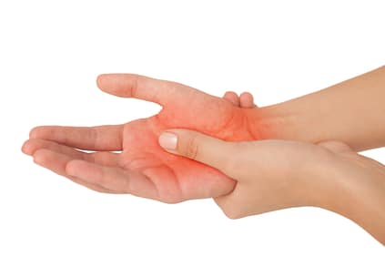

Source: wavebreakmedia, Standard License, via Shutterstock
Common Injuries
Learn more about common wrist injuries such as fractures, dislocations, and bursitis in our Common Injuries section.
Wrist Injury Diagnosis
The diagnosis of wrist injuries is remarkably diverse. A wide variety of tests are used to test, diagnose, and confirm these injuries. Apart from imaging, electromyography plays an important role in identifying whether the nerves or muscles are injured. Nerve conduction studies also confirm which part of the nerves are damaged.
Injury Specific Diagnosis
Physical Exam
On the physical exam, the doctor will observe the thumb, 2nd and 3rd digit, and the radial half of the 4th digit. They will check for the "flick sign." This is a sign where the symptoms improve when the fighter flicks their hand and wrist. The clinical physical exam also tests for sensory and motor deficits. Any evidence of thenar wasting is examined. There are several special tests specific to carpal tunnel syndrome. The best one is the carpal compression test. The doctor applies firm pressure directly over the carpal tunnel for thirty seconds. If the fighter experiences paresthesia or pain, the test is considered positive.
Phalen's test, the ‘reverse prayer,’ is also used. The doctor asks the fighter to fully flex both wrists by holding the backs of the hands together for about a minute. It is positive if that brings on tingling, pins and needles, or numbness in the thumb, index, middle and the thumb side of the ring finger — the median nerve's patch. Pain on its own does not count; it is too non-specific. The reverse version, palms together like praying, holds the wrists extended for a minute and is read the same way. Another test is the Hoffmann-Tinel sign: the doctor taps over the carpal tunnel, and tingling shooting into those same fingers is what makes it positive.
Imaging
X-rays are the first test ordered to review any past injuries, arthritis, or recent trauma. Ultrasounds and MRI’s can provide a three-dimensional view of the median nerve. However, electromyography and nerve conduction studies are diagnostic for Carpal tunnel syndrome. An electromyogram studies the functioning of the nerves and muscles by measuring the electrical impulses. These impulses are evaluated along the nerves, nerve roots, and muscles. It’s done by inserting a tiny needle electrode through the skin into the muscle. The electrode measures the electric impulses generated by muscle cells as they are activated by the nerves.
Nerve conduction studies measure how fast an electric impulse is traveling through a nerve. During a nerve conduction test, the nerve is stimulated. Its response is measured. If the response is slow in an area, that part of the nerve is compressed. In carpal tunnel syndrome, the electrical response is slower in the carpal tunnel.
Lab Tests
There are no blood tests to confirm Carpal Tunnel Syndrome. It is diagnosed clinically. Doctors may request other blood tests to confirm inflammation, but this is less likely.
To learn how Carpal Tunnel is treated Click Here
Physical Exam
On physical exam, the ganglion cysts are firm, well-circumscribed, and freely mobile masses. This is important as it needs to be differentiated from a sebaceous cyst or other inflammatory cysts. They are approximately 1-3 cm in size. Very often, they are fixed to the deeper tissue and not beneath the skin. The carpal tunnel, median nerve, and flexor tendon sheath are checked to see for carpal tunnel syndrome. The muscle innervation of the ulnar nerve is confirmed, and radial pulses are examined. This is because volar wrist ganglion cysts can cause ulnar nerve neuropraxia and compress the radial artery. This can lead to wrist ischemia. Occasionally, doctors will use the transillumination test. Here the doctor will dim the lights and use a bright light to illuminate the cyst and see if it is clear or contains material. Ganglion cysts usually transilluminate in this test.
Imaging
Plain films are ordered to rule out intraosseous causes. For ganglion cysts, radiographs are unremarkable. If the doctor wants to rule out a solid tumor, MRI is indicated. The MRI will show a well-circumscribed area. Ultrasound is also helpful, as it differentiates a cyst from a vascular lesion. This can be potentially dangerous if the cyst is biopsied. The puncture of the radial artery in a vascular lesion via needle aspiration can lead to profuse bleeding.
Lab Tests
There are no blood tests to confirm ganglion cysts. Imaging is mostly diagnostic and blood tests are only required if surgical intervention is recommended.
To learn how Ganglion cysts are treated Click here
On physical examination, doctors will check for wrist swelling and tenderness over the lunate. They will examine the nerves and muscles in the area. A thorough musculoskeletal exam and neurological exam is done to confirm synovitis and any loss of grip strength. Wrist range of motion exams is performed.
Kienböck disease is both a clinical and imaging diagnosis. For imaging, CT and MRI are highly specific. However, MRI is the most sensitive and can pick up occult cases that are easily missed on plain film and X-Ray. MRI images vary with the extent of osteonecrosis and how far it has progressed. It’s helpful because it can also demonstrate the integrity of the articulating cartilage.
Plain films can miss the disease especially early on because there is no diffuse lunate sclerosis. Cystic changes are absent and articular surfaces have not yet collapsed. There is no carpal collapse, mid-carpal, and radio-carpal in the early stages. To aid surgical planning, CT is sensitive. It can detect subtle subchondral fractures, coronal lunate fractures if present, any fragmentation, carpal instability in the fingers, and the degree of trabecular disruption.
Nuclear Scintigraphy was an adjunct for diagnosing early disease. Since it’s non-specific, it’s been replaced by the MRI. It uses tracers of radioactive molecules to diagnose diseases captured by gamma rays.
Staging is very important for this disease because it influences what treatment and the prognosis of the injury.
The Lichtman Classification is used, based on radiographic and MRI imaging.
Stage 1: Normal Xray, lunate intensity changes on MRI.
Stage 2: Lunate sclerosis on Xray with or without fracture. The lunate bone has a normal shape.
Stage 3: Collapse of the lunate articulating surface.
A: with preserved carpal alignment and height.
B: with scaphoid flexion and loss of carpal height.
C: with associated coronal fracture
Stage 4: Stage 3B + radio-carpal or mid-carpal arthrosis.
Functional Staging uses contrast-enhanced MRI to assess bone perfusion and the extent of necrosis. It is done to guide surgical techniques to revascularize the bone.
Stage 1: Bone marrow ischemia and edema but a viable lunate
Stage 2: Partial necrosis. The viable portion of bone is usually distal
Stage 3: Complete necrosis
Kienböck disease does not need to be confirmed with blood tests. The disease is diagnosed and staged with the help of imaging. Blood tests are only required for surgical treatment as part of the pre-operative clearance.
To learn how Kienbock's disease is treated Click here
The physical examination of the wrist usually uses the “look, feel, move” pattern. Doctors will first look for a deformity. Deformities are usually not visible in scaphoid fractures unless there are other associated carpal fractures. They will palpate the distal radius and ulna and metacarpals. This is to assess associated injuries and fractures. The classic sign is tenderness in the anatomic snuffbox.
This snuffbox is located between the first and third extensor compartments. Two muscles lie radially; abductor pollicis longus and extensor pollicis brevis. On the ulnar side, lies the extensor pollicis longus. Some tenderness is observed directly over the scaphoid tubercle. This is not always the case. The scaphoid tubercle is felt as a bony prominence at the level of the distal palmar crease.
Tenderness found in one of the three locations is clinically diagnostic:
Another more sensitive test is the scaphoid compression test. Here the doctor places the examining index finger and thumb over both poles of the scaphoid and compresses. If a fracture exists, then it will elicit pain. Typically, any pain in the anatomical snuffbox on the ulnar deviation of the wrist is suggestive of a scaphoid fracture.
Plain films are the first step in imaging the scaphoid fractures. AP and lateral views are taken. A special scaphoid view where the wrist is extended at 30⁰ and 20⁰ of ulnar deviation is done. Scaphoid fractures are easily missed on plain film. Up to 25% of scaphoid fractures are not visible on initial X-rays. Doctors might immobilize the wrist in a splint or cast for 1-2 weeks and repeat the Xray. For many fighters, bone scans are done to diagnose occult fractures 72 hours after the initial injury.
If symptoms persist and X-rays are clear MRI or CT is done. MRI is the most sensitive. It can also identify any injury to the ligaments or bone edema. CT scan is also fairly accurate but can miss fractures with less than 1 mm displacement.
There are no special laboratory tests unless surgery is to be done. For preoperative clearance, the routine blood tests, including full blood count, urea, and electrolytes, clotting screen and group and typing, are done.
To learn how Fracture of the Scaphoid are treated Click here


Common Diagnoses
Refer to our Common Diagnoses section to learn more about how injuries affecting the many ligaments and bones in the wrist joint are diagnosed.
Wrist Injury Treatment
The treatment of wrist injuries is diverse. It depends on how early the diagnosis is made and on the type of injury. Plenty of wrist fractures are treated in a cast rather than in an operating room, with surgery kept for fractures that are displaced, unstable, or failing to unite. Carpal tunnel syndrome often settles with conservative care, but severe or persistent cases do need surgery.
Injury Specific Treatment
Emergency Treatment
Carpal tunnel syndrome is rarely an emergency, since it builds up over time. There is one exception worth knowing: if numbness and severe pain come on suddenly after a wrist injury or fracture, that can be acute carpal tunnel syndrome caused by swelling inside the tunnel, and it needs urgent assessment at an emergency department rather than a night splint and a wait.
Medical Treatment
Conservative treatment is recommended for all fighters if carpal tunnel syndrome is identified early. The first step in treatment is to modify wrist activities that increase pressure in the carpal tunnel. Minimize repetitive flexion, extension, abduction, and adduction. For fighters who are bulkier, some weight loss can help. Combined therapy with wrists splints and physical therapy is better than single treatment alone. Pain medications like nonsteroidal anti-inflammatory drugs relieve pain. Though this is just a temporary solution.
Occasionally, if conservative treatment fails, a corticosteroid injection into the carpal tunnel is the next option, and an oral glucocorticoid can also give relief — usually temporary. Where symptoms are severe or keep coming back, surgery with carpal tunnel release is offered. It is not held back until nerve studies show heavy nerve damage: constant numbness, weakness, wasting of the thumb muscles or dropping things means the nerve is already suffering, and decompressing it late risks permanent loss. The procedure is minor and can be done endoscopically. The transverse carpal ligament, or flexor retinaculum, is divided to give the median nerve more room. It is typically a day procedure.
Home Treatment
Night wrist splints are recommended. Occupational hand therapy is beneficial. Many wrist issues may be helped by certain hand exercises or movements found in martial arts such as tai chi. See Fighting Arts article on “Body Strengthening Methods.”
Emergency Treatment
Ganglion cysts are usually not medical emergencies given that they take place over time and continuous micro-trauma. Asymptomatic lesions can be left as they are. Lesions that cause severe pain, weakness, or neuropathies must be treated.
Medical Treatment
Fighters who are asymptomatic can be observed, and many ganglion cysts go away on their own. Treatment also depends on the location of the cyst. Dorsal wrist ganglion cysts can be aspirated — a clinician draws the fluid off with a syringe under sterile conditions. Never try to drain, burst or hit one yourself: the radial artery, the tendons and the nerves all sit within millimeters of it, and you risk bleeding, infection and lasting damage. Cysts often recur after aspiration, so surgery is preferred where one keeps coming back.
Volar wrist ganglion cysts are not aspirated since they lie very close to the radial artery. The chances of bleeding are very high. Surgery is better for them and also for those fighters who are symptomatic. It’s also preferred in whom conservative treatment has failed. During surgery, the cyst is excised. An incision is usually made directly over the cyst. It is carefully dissected to expose the pedicle of the cyst. The capsule and all its attachments are removed to prevent any recurrence.
Home Treatment
Despite surgical excision, ganglion cysts have a high recurrence rate. Avoiding repetitive wrist motions for 6 weeks can help. Warm compresses improve circulation. Wrist braces and splints are also helpful.
If Kienbock's disease is suspected, then pain relief and immobilization is the first step. Seek a referral to an orthopedic doctor.
The goal of treatment in Kienböck disease is to relieve pain, preserve wrist motion, and preserve grip strength.
The treatment as mentioned earlier depends on the stage and the cause.
Stage I is usually managed first with splinting or casting and a change of training load.
Stage II is also treated with immobilization if the bone is not completely necrosed.
Stages II with complete necrosis, III, and IV usually require surgery. It is often a "joint-leveling" procedure, sometimes with bone grafting or the transfer of arterial branches from adjacent vessels.
Stages with lunate collapse and wrist degenerative changes may need more surgery like proximal row carpectomy. For those with shortened ulnas, the most common procedure to relieve the lunate is radial shortening osteotomy.
Following surgery, six to eight weeks of rest is needed. No immediate return to repetitive and wrist action is permitted.
Following any trauma to the wrist, if a scaphoid fracture is suspected, imaging must be done immediately. In suspected fractures with positive clinical signs on examination but no radiographic proof, imaging must be repeated in a week after splinting. If the pain persists and X-rays are still normal, then MRI or CT is advised. Immobilization with pain relief is advised.
The non-operative management or conservative treatment is advised for non-displaced fractures. It is also recommended for fractures within the distal third of the bone. The wrist is immobilized either with a long or short arm cast and with or without thumb spica to immobilize the thumb. The cast is kept for six weeks. X-rays are done after this to ensure the union of both parts of the scaphoid bone. The location of the fracture determines the time it takes to heal. Fractures in the distal-third heal within 6-8 weeks. Fractures in the middle-third heal in 8-12 weeks. Fractures in the proximal third take the longest; 12-24 weeks.
Surgical fixation is done when the displacement if more than 1mm, there’s a humpback deformity, the radiolunate angle is more than 15,transcaphoid perilunate dislocation, comminuted fractures, fractures in the proximal pole, non-displaced waist fractures in those in the combat arts who use wrist maneuvers, and those with nonunion or avascular necrosis after conservative treatment has failed.
During surgery, single or multiple screws are inserted. The procedure is done percutaneously or via open surgery. Open surgery is recommended for non-unions and large displacement. Percutaneous surgery is for acute, minimally displaced fractures.
For those fighters who continue to have nonunion, open reduction and internal fixation with bone graft are recommended. A bone graft is taken from the distal radius or the iliac crest.
Alternative treatments are salvage procedures, like excising the proximal fragment, total or partial fusion, radial styloidectomy, or proximal row carpectomy.
Immobilization is important as the scaphoid bone takes very long to heal. Any repeat injury can cause the bone to necrose forever as it fails to unite. Pain relief can be achieved with NSAIDs. Repeat X-rays at regular intervals are necessary to ensure the bone is healing.

Source: Vadym Huzhva, Standard License, via Shutterstock
Common Treatments
Learn more about how healthcare professionals treat injuries using physical therapy, medications, surgery, and other approaches in our Common Treatments section.