Combat Arts Thigh Injuries Overview
Leg kicks and oblique kicks in MMA can be powerful. They pack a punch. Josh Emmet suffered a knee injury in the early rounds against Shane Burgos. The ACL tear was just one of the injuries he suffered. He also had an impact fracture of the femur and it has put him on a long road to rehab. He refuses to quit.
Thigh muscles consist of the hamstrings at the back, the adductors on the inside, and the quadriceps at the front. The hamstrings extend the hip and bend the knee, the quadriceps straighten the knee (the rectus femoris also helping to flex the hip), and the adductors pull the leg toward the midline. Together they are the primary muscles used for balance and locomotion. Thigh injuries occur when one or more of these muscles is strained, torn, or bruised.
Thigh Injury Causes

It is worth being clear about what these muscles actually do. The hamstrings at the back of the thigh extend the hip and bend the knee; the quadriceps at the front straighten the knee, and only the rectus femoris also helps flex the hip; the adductors on the inside draw the leg toward the midline. Chronic hamstring injuries occur because a prior injury was improperly rehabilitated and small amounts of trauma are repeated over a long period (i.e., kicking or leg locks). Poor hamstring strength and flexibility increase the chance of small tears, which can leave the muscle tight and sore. A vicious circle is then established.
Thigh Injury Symptoms
Types of Thigh Injuries
Thigh injuries are a modest share of the injuries recorded in MMA injury surveys. Other than muscle contusions and lacerations, the most important injury in the thigh is that to the femur. The femur is one of the strongest and longest bones in the body. In striking sports the most common serious thigh injury is a deep quadriceps contusion from blocking or absorbing leg kicks: if that thigh keeps swelling and becomes tight, tense, and far more painful than the blow seemed to warrant, it needs emergency assessment for compartment syndrome rather than another ice pack.
Related Injuries
Femoral Fractures
Fractures of the femur can be life-threatening. High energy trauma can affect the neck and shaft of the femur. Powerful kicks to the thigh area can damage the bone. Stress fractures can also develop due to repetitive trauma and strain on the thigh. The femur is surrounded by three large muscle compartments that are susceptible to injury and hemorrhage.


Moreover, any injury to the femur could damage the sciatic nerve which lies very close to the bone. The femoral artery is also vulnerable to injury. It is the main artery of the whole lower limb, supplying the thigh as well as the leg below it. The risk of bleeding and shock from an injury to this artery is high. Since the femur is a large bone, there is also the risk of fat embolism as fat from the bone marrow leaks into the systemic circulation. A broken femur is therefore treated as a serious injury, and anyone who has one needs to be watched closely.
Femoral Fractures Causes
In combat sports, repetitive oblique kicks can cause stress fractures in the femoral neck and shaft. Acute injuries due to violent high-intensity punches, kicks, and sudden falls. Outside leg kicks with massive torque can injure the muscles and disrupt the bone. Similarly, high kicks with a full power leg can cause the neck of the femur to fracture.
Femoral Fractures Symptoms
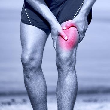

Source: Maridav, Standard License, via Shutterstock
To learn how Femoral Fractures is diagnosed Click Here.
Common Injuries
Learn more about common thigh injuries such as lacerations and muscle in our Common Injuries section.
Thigh Injury Diagnosis
To diagnose injuries in the thigh, the physical exam is important, but imaging is the key. This includes multiple modalities including plain films, CT, and MRI to evaluate the muscle compartments in the thigh. Moreover, since the femoral artery could be injured, to prevent hemodynamic compromise blood tests are also necessary to understand the extent of the damage.
Want to Know What to Expect When a Physician Diagnoses a Thigh Injury?
Get Our Free Newsletter to Get the Latest Injury Management Information.
Injury Specific Diagnosis
Femoral Fractures
Physical Exam
In trauma to the femur, the physical exam is done very quickly. The range of motion exercises of the hip and knee are done to rule out any injury to these joints. A very thorough neurovascular examination is done. Those with displaced fractures have a shortened and externally rotated lower limb. The ATLS (Advanced Trauma Life Support) protocol is used for femoral fractures, so vital signs are assessed and closely monitored. In the case of a fall, the ankle is also examined.

Source: Atstock Productions, Standard License, via Shutterstock
Imaging
Plain films must include the following views: anterior-posterior (AP) pelvis, AP and lateral hip, AP and lateral femur, AP and lateral knee. All bones and joints related to the thigh have to be assessed. The CT scan classifies the pattern of the fracture and can even visualize a subtle fracture line. It can also display and evaluate the femoral neck.
MRI is not used in an acute injury. For femoral neck stress fractures, they are better than the CT.
A chest radiograph and electrocardiogram (EKG) are necessary to evaluate the lungs and heart. This is to watch out for any signs of fat embolism and hemodynamic compromise.
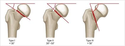
A femoral neck fracture is classified by Garden and Pauwel classifications as follows
The Garden Classification:
Type I: Incomplete fracture - non displaced
Type II: Complete fracture - nondisplaced
Type III: Complete fracture - partial displaced
Type IV: Complete fracture - fully displaced
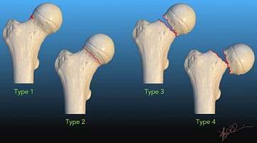
The type of fracture determines the treatment.
Pauwel Classification:
Type I: < 30 degrees
Type II: 30-50 degrees
Type III: > 50 degrees
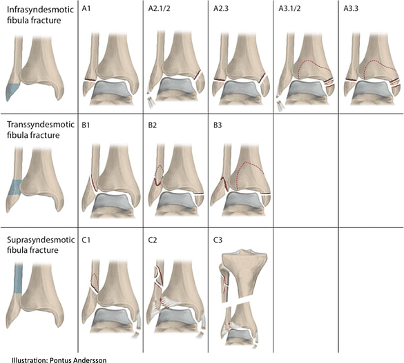
Classification of femoral shaft fractures by the AO/Orthopaedic Trauma Association classification. It classifies the fracture type resulting in 27 different patterns.
32A – Simple
A1 – Spiral
A2 – Oblique, angle > 30 degrees
A3 – Transverse, angle < 30 degrees
32B – Wedge
B1 – Spiral wedge
B2 – Bending wedge
B3 – Fragmented wedge
32C – Complex
C1 – Spiral
C2 – Segmental
C3 – Irregular
Lab Tests
Blood tests are done to assess hemodynamic status. This includes complete blood, count, basic metabolic panel, Rh, and typing for a cross match. In preparation for surgery, other blood tests like a coagulation profile might also be done.
To learn how Femoral Fractures is treated Click Here
Common Diagnoses
Learn more about how physicians use a variety of tests to diagnose injuries in the thigh in our Common Diagnoses section.
Thigh Injury Treatment
Femoral neck and shaft fractures are almost always treated surgically, especially in the acute setting. The alarming mortality figures often quoted for this injury come from studies of hip fractures in elderly patients, where the deaths are driven by age and other illnesses rather than by the break itself; they do not describe a young combat athlete with a broken femur. What is true for everyone is that a femoral fracture can bleed heavily and needs to be treated promptly.
Injury Specific Treatment
Femoral Fractures
Emergency Treatment
If a femur breaks in the gym or the cage, this is a call-an-ambulance injury. Do not move the fighter, and do not try to straighten or realign the leg. Keep them still and warm, control any external bleeding with firm direct pressure, give nothing to eat or drink, and watch for signs of shock — pale clammy skin, fast breathing, confusion — while you wait for the paramedics. In hospital, the Advanced Trauma Life Support protocol is activated since the thigh can hold around 1.5 L of blood. Any vascular injury can contribute significantly to shock. Intravenous fluids and cardiac monitoring are essential.
Medical Treatment
Treatment of femoral shaft fractures can be medical or surgical. The most common treatment for those in combat sports is intramedullary nailing. Other operative techniques include plate osteosynthesis and external fixation.

In some cases, closed treatment with traction, splinting, and casting is tried temporarily.
Intramedullary Nailing is done in stable athletes within 24 to 48 hours. It allows early weight-bearing and is therefore preferred.
Antegrade Nailing describes where the nail goes in rather than when: it is introduced at the top of the femur, near the hip, and it is the usual approach for femoral shaft fractures.
Retrograde Nailing is done if there are associated femoral neck and tibial fractures.
Reaming is a technique of preparing the medullary canal using a surgical reamer.
Plate Osteosynthesis is done via Open reduction internal fixation (ORIF) techniques. ORIF is typically not used as a primary treatment of femoral shaft fractures. It is done when IM nail fixation is contraindicated.
Minimally invasive techniques such as minimally invasive plate osteosynthesis (MIPO) does not expose the fracture site and bridge plating can be done instead.
External fixation is a damage-control measure rather than the routine treatment for every open fracture. It is chosen for unstable or heavily contaminated injuries, for fighters too unwell for a long operation, and where there is extensive vascular injury. It is often converted to internal fixation once the fighter is stable.
Traction can serve as a temporary measure. The following traction devices are used: Thomas, Hare, Sager, Kendrick, CT-6, Donway, and Slishman splints for apparent femoral injuries. Longitudinal traction can stabilize the fracture site, gross length, alignment, and rotation. It can relieve pressure on the nerves and blood vessels. It can restrict bleeding by stabilizing the surrounding clot. They are switched for fiberglass or plaster splint or skin vs. skeletal traction in the hospital.
Skin traction or Bucks traction is applied via a boot attached to a counterweight.
In skeletal traction, a pin is placed through the bone to prevent the soft tissues from bearing the traction force. Pins are usually placed in the distal femur, proximal tibia, and calcaneus.
Home Treatment
After surgery, the hospital stay is long. To prevent systemic complications, serial blood tests are required to assess anemia, renal insufficiency, and other metabolic disorders. The fighter’s thigh is checked daily for compartment syndrome, wound healing, and neurovascular compromise. Compartment syndrome means pressure building inside a closed muscle compartment, and the warning signs are pain far out of proportion to the injury, a tight swollen thigh, numbness or tingling, and severe pain when the knee or hip is moved passively. It is a surgical emergency: go to the emergency department immediately rather than waiting to see whether it settles.
After surgery, physical and occupational therapy begins in the hospital itself. They are important to regain mobility and function. At home, these exercises must continue. Weight-bearing is permitted as tolerated.
Driving is only allowed when a person can bear weight and brake safely without any pain. Smoking is not permitted, as it slows bone healing.
Common Treatments
Our Common Treatments section discusses the various treatments for injuries to the thigh.