Combat Arts Shin Injuries Overview
In the second round of the UFC 168 Middleweight Championship fight between Anderson Silva and Chris Weidman in 2013, Anderson sustained a horrific injury while executing a leg kick against Weidman. The fight was stopped when Anderson broke his left tibia and fibula, requiring surgery to insert a titanium rod in his tibia.
Shin Injury Causes
Fractures of the tibia and fibula are the most common fractures in the lower extremity. Increasing loads can also stress the bones of the shin causing them to develop stress fractures. High-velocity kicks can break the tibia and fibula. A sudden blow can also cause the leg to buckle and the bone to break.
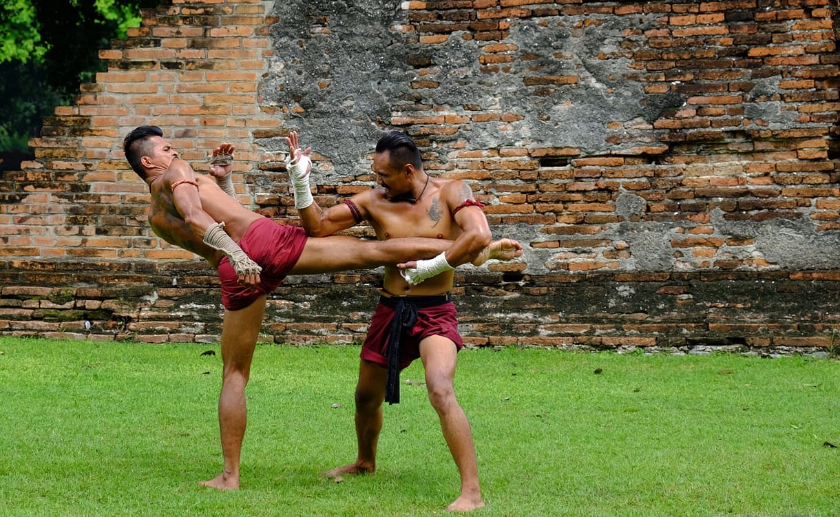
Source: Maliwan kittidacha, Standard License, via Shutterstock
Many martial artists avoid wearing shin guards or shin pads because they feel they slow them down, which leaves the bone taking repeated impact unprotected. Shin conditioning is an important part of Muay Thai for a different reason: there the shin is the weapon, and fighters load it gradually so it tolerates hard contact. Traction forces develop on the leg, either on the bone or the muscles, and cause various chronic injuries.
Shin Injury Symptoms

Types of Shin Injuries
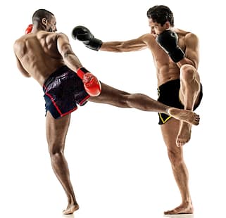
Since the tibia and fibula are an integral part of the shin, injuries to these bones are of primary concern. The injuries commonly seen in martial artists are fractures of the tibia and fibula, stress fractures, and medial tibial stress syndrome, better known as shin splints. Stress fractures and MTSS are not the same condition: MTSS is a bone-stress reaction along the inner border of the tibia, while a stress fracture is an actual crack in the bone. Untreated, MTSS can progress into one.
Related Injuries
Shin splints, or medial tibial stress syndrome, is pain along the inner border of the tibia where the muscles attach to the bone, involving the bone tissue itself as well as the overlying muscle and tendon. Repeated loading irritates the bone and its covering; it does not pull the muscles, tendons, and ligaments off the bone. Left unchecked, that bone stress can progress to a stress fracture, but most cases of shin splints do not.
Shin splints may be very mild creating only a simple stress response or bad enough to damage the shin bone with a severe stress fracture. Shin splints are one of the common causes of shin pain in martial artists.
Symptoms
Shin splints may occur on the front or anterior part of the shin bone or the inside rear or posterior part of the shin bone. The placement of injury is shown by where pain flares up during movement. If pain occurs when lifting the toes while keeping heels on the ground it is most likely from anterior shin splints, but if there is pain along the inside rear of your shin bone it is probably from posterior shin splints or tibia stress fractures.
Symptoms may include:
Causes
Shin splints occur from overuse and improper use of the leg. Shin splints and shin pain are common results of overloading, overtraining, training on inappropriate surfaces (cement, tile, etc.), and pushing the body too hard, too fast. A martial artist doing 400 roundhouse kicks in a row on a heavy bag, or training for Iron Leg without allowing the body sufficient recovery time, for instance, may develop shin splints.
Causes include:
To learn how Shin Splints is diagnosed Click Here
Overview
Brazilian UFC fighter Thiago Santos went through his entire bout with injuries. His loss was compounded by several of them: three torn ligaments, a torn meniscus, and a fractured tibia. That he kept fighting astonished his audience and his opponent, but it is not something to copy — carrying on with a broken tibia risks displacing the fracture and damaging the nerves and blood vessels around it, and the cost is paid in the months afterwards. Tibial fractures are common in athletes who use their legs to fight, train, or compete. Horrific tibial fractures have been seen among Olympic gymnasts, pole vaulters, runners, baseball players, combat artists, kickboxers, and others due to accidental falls, wrong landings, and sudden kicks. Even in sports where shin guards are used like soccer, tibial fractures are common and horrific. The injury is obvious because the deformity is clear to see. If there is an open fracture, the very sight of the bone jutting out can be distressing.
Symptoms
Causes
There are two ways to sustain a tibial fracture. One pattern is that of a lower energy force, the other is a high energy mechanism. In a low-energy injury, it is usually torsion that acts on the tibia. The torsional force from indirect trauma affects the tibia. These could be trauma to the thigh, femur, or fibula that causes spiral fractures as the forces travel down the tibia. Here there is less soft tissue injury and it could be associated with a fibular fracture as well. In high-energy injuries, there is direct trauma. Any high-velocity impact through a punch, kick or jab can cause a wedge or short oblique fracture. These fractures can be severe enough to fragment the bone and cause significant soft-tissue injury.
To learn how Tibial Fractures is diagnosed Click Here
Fibular fractures usually occur due to trauma and in concurrence with tibial fractures. That’s not always the case. Take, for instance, US Olympic taekwondo bronze medalist Steve Lopez. His hopes of adding a third Olympic title ended in his first-round fight. Since 2000, when taekwondo became a full Olympic sport, Lopez had always taken home a medal. That year was different. He fought on through pain that was eventually diagnosed as a broken right fibula. Take the opposite lesson from it: pain in the shin that keeps getting worse, or that you cannot walk on afterwards, should be imaged rather than trained through.
The fibula is one of the two long bones that form the shin in the leg. Unlike the tibia, it carries only a small share of the load passing through the leg, so it is much smaller and thinner and more easily broken. In contact sports, the fibula is rarely injured in isolation. Fibular injuries accompany injuries to the tibia as a result of a collision, falls, and high-velocity kicks. They get missed because they are often mistaken for an ankle sprain.
To learn how Fibular Fractures are diagnosed Click Here

Source: Amorn P, Standard License, via Shutterstock
Common Injuries
Our Common Injuries section has more about shin contusions and lacerations that are sustained during a bout or training.
Shin Injury Diagnosis
The diagnosis of shin injuries is typically through physical exams and imaging. It’s not hard to visualize fractures of the tibia and fibula since the deformities are visible to the naked eye. However, stress fractures require imaging for confirmation. Blood tests are only required in the case of repetitive stress fractures to rule out other metabolic diseases.
Injury Specific Diagnosis
Physical Exam
To diagnose medial tibial stress syndrome physical examination is extremely important. The fact that the martial artist has a long history of stress or overuse of the leg supports the diagnose of shin splints. The doctor will check for any exercise-induced pain along the distal two-thirds of the medial tibial border. If there is pain during activity or after any activity which reduces with rest, this a sign of MTSS. Any cramping or burning pain in the back of the leg is also checked. Doctors will test for numbness or tingling in the foot.
As they examine, they will inspect and palpate the entire lower extremity. Pain reproduced by pressing along the posteromedial border of the tibia, over a span of several centimetres rather than at one spot, points to MTSS. Findings such as severe swelling, redness, or absent pulses in the foot argue against shin splints. They are red flags for compartment syndrome, a blood clot, or a problem with the blood supply to the leg, and they call for urgent medical assessment rather than a diagnosis of MTSS.
Imaging
Medial tibial stress syndrome is a clinical diagnosis. Imaging is only done to rule out other injuries. Plain radiographs are usually normal, and early stress fractures often show nothing at all on plain film. The so-called dreaded black line is a late finding seen on the front of the tibia in some stress fractures: when it is there it matters, but its absence rules nothing out.
MRI is the best imaging modality for MTSS. It can also detect higher grade stress injuries in the bone such as a tibial stress fracture. Nuclear bone scans are another alternative. They are not as specific but can be used. On MRI, periosteal edema and bone marrow edema are seen.
Nuclear bone scans show radionuclide uptake in the cortical bone. This increased uptake has a characteristic “double stripe” pattern. High-resolution CT is another alternative but not as sensitive as an MRI.
Lab Tests
In martial artists who have recurrent stress fractures or develop MTSS frequently, vitamin D level in the blood is tested. Calcium, phosphate levels, and renal function tests are done.
To learn how Shin Splints is treated Click Here
Physical Exam
During the physical exam, the range of motion and examining the stability of the joint is virtually impossible due to the gross injury. In most cases, the neurovascular exam is done. This is to ensure that peripheral pulses and nerve supply are still intact. Doctors will palpate the dorsalis pedis and posterior tibialis arteries.
Doctors will also check the soft tissues for signs of compartment syndrome: tightness in the muscles, shiny skin, pain that is out of proportion to the injury, redness, numbness or tingling in the foot, and severe pain when the toes are passively stretched. Compartment syndrome happens when swelling inside the closed muscle compartments of the lower leg squeezes the blood supply. The exertional form builds up during training and eases with rest, and it is worth investigating if it keeps happening. The acute form follows a fracture or a heavy blow, keeps getting worse, and is a surgical emergency — that fighter belongs in the emergency department now, not in an ice bath waiting to see. The skin is also examined for lacerations, abrasions, fracture blisters, and the condition of the leg compartments.
Imaging
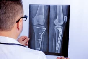
Plain films are initially obtained and usually diagnostic. The following views are obtained; full-length AP and lateral views of the affected area, AP, lateral and oblique views of the knee and ankle, the same side of the injury. CT scans can identify any joint involvement and rule out a posterior malleolar fracture. The latter is seen in spiral distal third fractures. If joint involvement is suspected a CT scan is done. Based on the imaging, the tibial fractures are classified via two systems. The first is Oestern and Tscherne. It classifies closed fractures.
Grade 0: Injuries by indirect forces, minimal soft tissue damage
Grade 1: Superficial contusion/ abrasion, simple fractures
Grade II: Deep abrasions, muscle/skin contusion, direct trauma, impending compartment syndrome
Grade III: Excessive skin contusion, crushed skin or muscle destruction, subcutaneous degloving, acute compartment syndrome, and rupture of a major blood vessel or nerve
The Gustilo-Anderson classifies open tibia fractures:
Type I: limited periosteal stripping, clean wound < 1 cm
Type II: mild to moderate periosteal stripping; wound > 1 cm
Type IIIA: significant soft tissue injury, significant periosteal stripping, wound > 1 cm, no flap required
Type IIIB: significant periosteal stripping, significant soft tissue injury, flap required
Type IIIC: significant soft tissue injury, a vascular injury requiring repair
Lab Tests
Blood tests are not necessary to diagnose fractures of the tibia.
To learn how Tibial Fractures are treated Click Here
On physical exam, the doctors will palpate the leg for tenderness. This is especially over the fibula on the side of the leg. Pain here could indicate a fibula shaft fracture. The skin is examined for lacerations, abrasions, contusions, bruising, and signs of compartment syndrome. The doctors will test the range of motion of the joints above and below the injury. Ligaments of the knee are also checked. This means testing the knee and ankle to rule out intra-articular injuries. A neurovascular exam is done to ensure the nerve supply is intact.
It is necessary to obtain weight-bearing anterior-posterior, lateral, and oblique films. Plain films of the knee and ankle are done to rule out fractures of other bones like the tibia. Imaging identifies simultaneous fractures. A fracture of the proximal fibula combined with a syndesmotic injury at the ankle is a Maisonneuve fracture — by definition it is never an isolated break, so a fracture high on the fibula should always raise suspicion of ligament damage down at the ankle. That changes the management plan for the injury. Similarly, any fibular fracture within 15 cm of the ankle joint can involve the tibia, ligaments, or both. A CT scan diagnoses rotational deformities and malunion of the syndesmosis.
Blood tests are not necessary to diagnose fibular fractures. Imaging and physical exam are diagnostic.
To learn how Fibular Fractures are treated Click Here

Source: MelenaSlavina, Standard License, via Shutterstock
Common Diagnoses
Browse through our Common Diagnoses section to learn more about how various imaging modalities are used to diagnose injuries to the tibia and fibula.
Shin Injury Treatment
The treatment of shin injuries depends on several conditions; type of injury, closed versus open, acute versus chronic, the involvement of other joints and ligaments, age of the fighter, and associated metabolic conditions.
Injury Specific Treatment
Emergency Treatment
Shin splints are not an emergency. The treatment is conservative and means relative rest: stop the activity that hurts, keep moving with low-impact work that stays pain-free, and build the loading back gradually. Bed rest and staying completely off the leg are not recommended — they cost conditioning and carry their own risks without speeding anything up.
Medical Treatment
MTSS treatment is usually rest and the modification of activity. Stop all repetitive, load-bearing exercise. The duration of rest depends entirely on the individual. Various add-on treatments are tried, with limited evidence behind any of them: cryotherapy or ice massage, iontophoresis, phonophoresis, ultrasound therapy, and extracorporeal shockwave therapy. Periosteal pecking is sometimes listed alongside these, but it is a surgical procedure, not a therapy to try at home.
Evidence for the add-ons is weak. Stretches, low-energy laser therapy, lower leg braces, and compression stockings have not been shown to do much on their own. Strengthening and gait retraining are a different matter — they are part of standard rehabilitation for shin splints, as below.
Some research suggests prefabricated orthotics may help prevent shin splints in certain groups, but the evidence is limited and is mostly about prevention rather than treating an existing case.
In martial artists who have MTSS repeatedly or those who respond rather slowly to conservative treatment with rest and modifying activity, it may help to optimize calcium and Vitamin D. Physical therapy must include gait retraining and strengthening exercises.
Severe tibial stress fractures require surgery if they do not respond to medical or conservative therapy.
Home Treatment
Most martial artists recover fully with adequate rest and by modifying their activities. Medial tibial stress syndrome can progress to a tibial stress fracture. This is because any cortical microtrauma can evolve into a larger cortical fracture. This is not the case with every MTSS injury.
MTSS is a stress reaction to the tibia due to overuse. It is therefore important for a martial artist to have a graded exercise regimen. Avoid overtraining. Allow adequate recovery time after each training. Optimize vitamin D and calcium levels through supplementation.
Emergency Treatment
An open fracture — bone visible through broken skin — is time-critical. At the scene: call emergency services, cover the wound with a clean dressing, never push the bone back in and never wash the wound out, support the leg in the position you found it, and get the fighter to hospital. Antibiotics are given as early as possible for an open fracture and tetanus cover is checked. After initial trauma protocols are followed, doctors perform wound debridement as the primary emergency procedure, without a planned second operation, for open tibial fractures.
Medical Treatment
Non-operative treatment is chosen for closed fractures. A long leg cast that offers closed reduction is preferred for fractures with an angulation of fewer than 5 degrees of varus-valgus. Closed reduction is also preferred where the anterior-posterior angulation is less than 10⁰. If there is more than 50% cortical apposition, less than 1-cm shortening, less than 10- 20 degrees of flexion, and less than 10 degrees of rotational malalignment after reduction, then closed reduction is done.
Surgical treatment includes many techniques. The first is external fixation. It is preferred when there is significant soft tissue injury. If the neurovascular status is compromised, then this is the go-to procedure. Intramedullary Nailing (IMN) is chosen for fixation via surgery as it is associated with less malalignment, the time taken for the union of the bone is less and the time to weight bearing is much faster.
Percutaneous Plating-Shaft is used in fractures of the distal tibia and proximal third fractures. These fractures are too close or too far for IMN.
Amputation is another option, although not many combat fighters will want it. If the leg is severely mangled, scoring systems such as the mangled extremity severity score (MESS) are sometimes used to describe the damage. They have repeatedly proved poor at predicting which limbs can be saved, so no score on its own decides an amputation. Significant soft tissue trauma, warm ischemia for more than 6 hours, and associated foot trauma all weigh into that decision, which belongs to the surgical team and the fighter together.
Home Treatment
In martial artists, where the tibial shaft fractures are treated conservatively, the combat artist has to keep the affected leg in a long leg cast. He has to be non-weight bearing for 6 weeks. Those who have surgery like external fixation, especially unstable fractures will require longer periods of inactivity. These combat artists have to be non-weight bearing for 6 weeks for articular fractures outside the joint and for 12 weeks for intraarticular fractures.
In length-stable fractures, some doctors may allow patients to bear weight as tolerated. In cases where the tibial fractures were treated via IMN or plating the same applies as for external fixation. Tolerate weight and use pain as a guide as return to activity commences. During the recovery period, maintain active knee and ankle range of motion. DVT prophylaxis care is given to all athletes who are non-weight bearing when it comes to lower extremity fractures.
Most fibular fracture injuries are treated symptomatically. The treatment is conservative, with adequate pain management and protected weight-bearing — usually a walking boot rather than a knee brace. Early knee and ankle motion is encouraged to prevent joint stiffness.
No stabilization is required for proximal fibular fracture in a Maisonneuve injury. Intraosseous and or syndesmotic injury require fixation near the ankle. This is the case if the distal tibiofibular joint is highly unstable. Closed reduction and casting are an alternative treatment.
Open reduction and internal fixation are only done for acute injuries where there are avulsions of the LCL.
In most cases, isolated fibular shaft fractures are treated conservatively. This is possible as the fibular shaft is not a weight-bearing bone. Weight-bearing is slightly restricted and martial artists are mobile with a walking boot.
Isolated fibula shaft fracture typically requires only symptomatic treatment. For most athletes it leaves no long-term limitation, and the bone generally unites over a couple of months, though healing times vary with the fracture and the fighter.
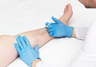

Source: HenadziPechan, Standard License, via Shutterstock
Common Treatments
Read more about treating injuries in the shin in our Common Treatments section.
Featured Image Source: Magic mine, Standard License, via Shutterstock