Combat Arts Eye Injuries Overview
Combat sports, martial arts, and fight events have a high prevalence of eye injuries. Glove design has been revised over the years partly to cut down on eye pokes, and open-fingered gloves remain a recognised hazard rather than a solved problem. Torn eyelids and retinal damage are other common injuries.
Eyes are delicate and require immediate intervention if an injury occurs, dirt, hair, or other foreign material are present, infection or other physical issues arise. It is important to recognize danger signs when looking at the eyes and to understand the proper response.
Eye Injury Causes
Former UFC middleweight champion Michael Bisping almost lost the sight in one eye after he kept fighting with a detached retina. He kept winning, and he ended up with permanent damage to that eye. Read that as the warning it is, not the war story it sounds like: a detached retina does not heal by being ignored, and every round fought on one makes the surgeon’s job harder and the final vision worse. Bisping’s career began with winning “The Ultimate Fighter 3,” and he finally hung up his gloves when ongoing vision problems convinced him to quit. Fans poured out the love when he said no to one last round in the cage.

A strike to the eye, finger poke, face plant into the mat, gi material, hair or other substance rubbed into the eye can cause irritation or damage.
Eye Injuries Symptoms
Types of Eye Injuries
8.3% of all injuries in martial arts involve the eye. Eye injuries for mixed martial artists fall into three categories: Abrasions, penetrating injuries, and blunt trauma. These include: Corneal Abrasion, Retinal Tears, Ocular Fractures and Penetrating Injuries.
Don't Lose Sight of Your Ultimate Fitness Goals!
Let Us Help You Prevent and Treat Eye Injuries.
Related Injuries
The most common eye injury in martial arts and combat sports is corneal abrasion or a scratch on the eye. The cornea is the outermost structure covering the eye. Although these injuries normally heal quickly and create no long-term harm, they can be extremely painful. Deeper abrasions may cause scarring on the eye, which could create vision problems.
“They say it is worse than a kidney stone,” says Dr. Pang of corneal abrasions. “It’s a sharp, painful like a knife going into your eye, but it is really pretty minor unless it gets infected. The recovery is about 3-4 days usually.
Symptoms
To learn how a Corneal Abrasion is diagnosed, click here.
The retina is a very thin and delicate membrane containing the rods and cones required to see color, light, and dark. If that thin layer receives damage, the entire retina can detach over time. Fighters have gone blind because of this.
“A retinal detachment occurs when the lining of the eye comes off. You can think of the retina like an egg. You have an egg and you take the egg out, the whites and the yolk and then there is the very lining of the eggshell. If you have a tear in the retina it can be treated with a laser or freezing of the tear and it will scar down.
If you have an undiagnosed tear or hole in the retina fluid can go underneath it and cause it to detach. If fluid from within the eye passes through a retinal tear in it can cause the separation of the retina from its underlying tissue resulting in detachment,” says Dr. Pang.
“The only reason to stop a fight because of eye injuries would be a big laceration causing bleeding to the eye, when the fighter states he can’t see anymore, or if there is an orbital fracture,” said ophthalmologist Dr. Pierre Pang, M.D., an eye and retinal specialist who has served as the official doctor at many MMA events on Oahu. That is one ringside physician’s threshold for stopping a bout, and it sits well below the danger signs listed further up this page. Blood in the clear part of the eye, a pupil that is uneven or oddly shaped, a sudden shower of floaters, flashes of light, or a curtain moving across the vision are all reasons to stop and have the eye examined. None of them looks dramatic from outside the cage, and every one of them can end in permanent vision loss if the fighter carries on.
Symptoms
Causes
There are three types of retinal detachment. Rhegmatogenous detachment is the one that matters most to fighters: a tear or hole opens in the retina, and fluid from inside the eye passes through it and lifts the retina off the tissue beneath. Blunt trauma is one cause of that tear, and age-related changes in the vitreous jelly are another — which is why it can also happen to an older martial artist with no blow to the eye at all.
Tractional detachment happens when scar tissue on the surface of the retina contracts and slowly pulls it away from the back of the eye. In practice it is driven far more by advanced diabetic eye disease than by fight trauma. Exudative detachment is the least relevant of the three to a fighter: fluid collects under an intact retina because of inflammation, vascular disease, or a tumour, with no tear involved at all.
To learn how Retinal Tears are diagnosed, click here.
MMA veteran light heavyweight Brandon Vera knows what it means to have an orbital fracture. He suffered a tripod orbital fracture when a piece of bone went into his eye. He was elbowed in the face. In martial arts, while unintentional, it’s easy to catch someone’s arm or foot in the face. The blunt force can cause orbital bones to fracture.
Symptoms
Causes
A martial artist can take an orbital rim fracture, where the lower rim over the cheekbone or the upper rim below the forehead is broken. They can also take a blowout fracture, an indirect orbital fracture: when the eye socket catches a fist or a foot, the bony rim stays intact but the paper-thin floor of the orbit gives way underneath the eye. That is a break in the bony socket. It is not a rupture of the eyeball — a ruptured globe is a different and far more serious injury.
Direct orbital fractures are those where both the bony rim and the floor of the socket are broken.
To learn how Ocular Fractures are diagnosed, click here.
Penetrating eye injuries are common in martial arts because no one wears any protective eye equipment. These injuries occur when something cuts your eye. It could be a weapon, corrective glasses, or even someone’s fingernail.
Symptoms
Causes
A penetrating injury needs a sharp object — a fingernail, a broken pair of glasses, a weapon. A fist delivers blunt force, and blunt force to the eye causes bruising, blood in the front chamber, an orbital fracture, or in severe cases a ruptured globe, rather than a penetrating wound. Both routes are common in martial arts because nobody wears eye protection, and both can cost a fighter their sight.
To learn how Penetrating Injuries are diagnosed, click here.
A hyphema is bleeding into the front chamber of the eye, the space between the clear cornea and the coloured iris. In combat sports it is usually caused by a blunt blow — a punch, kick, elbow or knee. A small hyphema may only be visible to a doctor with a slit lamp; a larger one shows as a layer of blood at the bottom of the iris or can fill the front of the eye.
Symptoms
Blurred or reduced vision, eye pain, sensitivity to light, and blood visible in front of the iris or pupil. Nausea and vomiting can mean the pressure inside the eye has risen sharply.
Causes
A blunt impact tears small blood vessels in the iris or at the drainage angle of the eye. The same blow often causes other damage — an orbital fracture, a torn iris, a dislocated lens or a retinal injury — which is why every hyphema needs an eye specialist. People with sickle cell disease or trait, or who take blood-thinning medication, are at higher risk of complications.
To learn how Hyphema is diagnosed, click here.
Common Eye Injuries
Find out more about common injuries such as lacerations and facial fractures that can also affect the orbital area in our Common Injuries section.
Eye Injury Diagnosis
The first step in diagnosing eye injuries is the ophthalmic exam. This tests pupillary light reflex, red reflex, eye movement, and nerve functions. Following this, the doctor will use an ophthalmoscope to look into the eyes. They might even request fluoroscopy and order imaging to study the blood vessels and the bones of the orbit.
Injury Specific Diagnosis
Physical Exam
The ophthalmic exam will usually display extreme sensitivity to light, excessive tearing, and decreased visual acuity.

Source: Ground Picture, Standard License, via Shutterstock
To facilitate the exam, a topical anesthetic is used. Extraocular movements are examined and documented.
Imaging
Lab Tests
To learn how a Corneal Abrasion is treated, click here.
Physical Exam
Regular eye exams pick up retinal tears before they progress, and that is the point of having them. A tear does not heal on its own: it is sealed with laser or freezing treatment so fluid cannot track underneath it, and sealing it is what prevents a detachment. Fighters who take repeated head and eye trauma should have a dilated retinal examination on a regular schedule, not only after an obvious injury.
Usually, when a retinal tear is suspected, visual acuity at near and distance is checked. The external examination will involve searching for signs of trauma. The ophthalmologist will also check the visual fields through a confrontation examination to isolate the location of the retinal detachment. The ophthalmoscopic exam will diagnose a retinal detachment fairly accurately.
Imaging
In most cases, an ophthalmoscopic examination is enough. However, if foreign body insertion is suspected or the doctor wants to see the real-time movement of the detached retina, an ultrasound might be advised. CT imaging can visualize folded membranes with subretinal space fluid.
Lab Tests
Blood tests like HbA1c and CBC are done to determine any underlying causes for retinal detachment like diabetic retinopathy and sickle cell disease. Since surgical intervention will be required, baseline blood tests like BMP, PT/PTT, and INR, are useful.
To learn how Retinal Tears are treated, click here.
Physical Exam
The doctor will test the extraocular movement in each eye separately, as well as confrontational visual fields. They will also do a swinging-flashlight test to rule out afferent pupillary defects, color plates, and red desaturation. While painful, they will palpate the bony rim and check extraocular movements.
Imaging
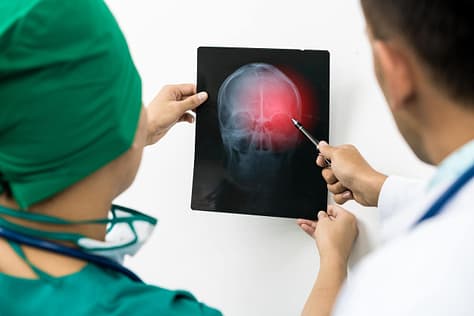
CT ‘s and MRI’s are important for detecting fractures and soft tissue injuries when it comes to the orbit.
Source: Summit Art Creations, Standard License, via Shutterstock
Lab Tests
Blood tests are usually not required unless other signs of trauma are noted and a high risk of infection is suspected.
To learn how Ocular Fractures are treated, click here.
Physical Exam
The physical exam here must include palpating the orbital rim and checking for any orbital fracture. Doctors will check for any foreign body. The eyes are quickly examined without putting any pressure on the globe with the help of Desmarres retractors.
Imaging
Imaging is usually with plain film x-rays of the head and/or CT scans. These will determine the location of the injury and the presence of a foreign body if any. A CT scan will also demonstrate the shape and integrity of the traumatized globe.
Lab Tests
Blood tests are required only if surgical intervention is recommended.
To learn how Penetrating Injuries are treated, click here.
Physical Exam
An eye doctor checks vision, measures the pressure inside the eye, and examines the front of the eye with a slit lamp to grade how much of the chamber is filled with blood. They also look for other damage from the same blow, including the lens, the retina and the bones around the eye.
Imaging
If the back of the eye cannot be seen, or a fracture or ruptured globe is suspected, a CT scan of the orbits is done. Ultrasound of the eye is avoided until a ruptured globe has been ruled out, because the probe presses on the eye.
Lab Tests
Sickle cell testing is recommended when a person’s status is unknown, because sickle cell disease or trait raises the risk of dangerous pressure rises and optic nerve damage. Clotting tests may be done if a bleeding disorder is suspected.
To learn how Hyphema is treated, click here.
Diagnoses of Common Eye Injuries
To learn more about common eye diagnoses visit our Common Diagnoses page.
Eye Injury Treatment
In general, eye injuries need special care. They are common in mixed martial arts and contact sports where no eye equipment is used. Not all ocular injuries will require surgical treatment.
The key is getting medical attention urgently whenever an eye injury is suspected, and following a few basic rules until you get there. Any eye injury with a change in vision, real pain, blood visible in the clear part of the eye, or a pupil that is no longer round and equal needs same-day ophthalmology assessment. That includes hyphema — blood pooling in the front chamber of the eye — which is one of the more common serious eye injuries in combat sports and carries a risk of re-bleeding and of raised pressure inside the eye. Never rub an injured eye, never press on it, and never try to pull out anything embedded in it.
Injury Specific Treatment
These steps apply to any eye injury in the gym or ring. When in doubt, protect the eye and get it seen the same day.
Particle, Sand, or Foreign Material in Eye
- Never rub the eye
- Lift the upper eyelid gently and let tears rinse the eye, or flush it with clean water, saline or an eye wash made for the purpose
- If the particle won’t rinse out, close the eye or cover it lightly and get emergency care
- Do not try to remove an object that is stuck in the eye or under the eyelid
- Do not put antibiotic or other medication in or on the eye unless a doctor prescribes it
In Case of a Cut or Punctured Eye
- Never rub or press on the eye, and do not try to remove an object stuck in it
- Do not rinse the eye with water
- Protect the eye with a rigid shield or the bottom of a paper cup taped over it, without touching the eye
- Do not give aspirin, ibuprofen or other anti-inflammatory pain drugs, which can increase bleeding
- Go to the emergency room or an eye specialist immediately
Chemical Burn
Start flushing the eye with clean water or saline immediately and keep flushing continuously for at least 15 to 20 minutes. Hold the lids apart and let the water run across the eye. Do not stop early to go looking for help — have someone else call while you keep irrigating, and keep irrigating on the way to the emergency department.
In Case of a Blow to the Eye
- Never apply pressure to the eye
- Hold a cold pack very gently against the cheek and brow, not pressing on the eye, to ease pain and swelling
- Get same-day medical care for any change in vision, blood visible in the eye, double vision, pain when moving the eye, numbness of the cheek, or a pupil that is not round and equal
Emergency
Before anything goes near the eye, rule out a penetrating injury or a ruptured globe. If the eye is cut or punctured, the globe looks distorted, the pupil is misshapen, or something is stuck in it, do not irrigate and do not press on it — shield the eye and go straight to an emergency department. For a simple surface abrasion, saline irrigation is usually the first step to flush out the grit. Never rub the eye. And never keep using the numbing drops a doctor used for the examination: they feel like relief, but they delay healing and mask an eye that is getting worse. If symptoms continue, seek prompt medical attention.
Medical
Emergency
A retinal tear that has not yet progressed to detachment is still urgent. It is normally sealed with laser or freezing treatment within days, not booked at leisure. If the retina is already detached it needs prompt surgery, and a detachment that has reached the centre of vision is treated as an emergency.
Medical
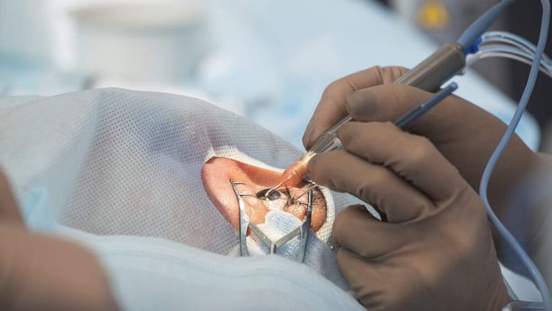
Retinal tears and detachments almost always need a procedure. Sealing a tear with laser photocoagulation can be done in the clinic. Repairing a detachment that has already happened is operating-theatre surgery under anaesthetic, which is a different undertaking entirely. For tears, laser surgery welds the retina down through photocoagulation.
Source: SERGEI PRIMAKOV, Standard License, via Shutterstock
Cryopexy — freezing the tear so it scars down — can also secure the retina. For detachments, surgeons can introduce intraocular gas to push the retina back and close any gaps. They can fit a band around the eye, called scleral buckle surgery, or drain the fluid inside the eye through a vitrectomy. To flatten the retina, air, silicone oil, or gas is injected into the vitreous space. Scleral buckle, vitrectomy, and gas or oil tamponade are all theatre operations, not clinic-room procedures.
Home
Following any retinal tear or detachment, martial artists are advised bed rest to avoid any activities that can further worsen the tear or cause a second detachment.
Emergency
Do not patch the eye. All orbital fractures require emergent care. If possible, place an eye shield. Avoid any drops or ointments.
Medical
Treatment depends on the locations and severity of the fractures. Small uncomplicated fractures will not require surgery. Severe surgeries might require reconstruction to remove bony fragments, free tapped eye muscles, repair deformities, and restore normal orbital architecture.
Home
Simple fractures are managed with ice packs, nasal decongestants, and antibiotics taken by mouth. An orbital floor fracture opens into the sinus, so antibiotic eye drops do not reach where the infection risk actually is. Rest, and do not blow your nose — that can force sinus air into the orbit.
Emergency
Do not force the eyelids open if they are closed and do not attempt to remove any protruding object from the eye. Doing so will cause extrusion of the ocular contents. Do not let the fighter eat anything.
Medical
Penetrating eye injuries require surgery. The eye is protected with an eye shield and systemic analgesics and antibiotics are administered.
Home
Following surgery, daily follow up with an ophthalmologist is advised to ensure the eye is healing well. Bed rest is recommended along with topical antibiotics to avoid bacterial super-infection.
Emergency
Stop the fight or training session and get same-day assessment by an eye specialist or emergency department. Protect the eye with a rigid shield or the bottom of a paper cup, never a pressure patch. Keep the head raised, and do not take aspirin or ibuprofen, which can make the bleeding worse.
Medical
Most hyphemas are treated without surgery: rest with the head raised, a protective shield, eye drops to calm inflammation and pain, and drops to lower the eye pressure if it rises. The eye is re-checked over the following days, because a second bleed, usually within the first week, is the main danger. Surgery to wash the blood out is needed if the pressure cannot be controlled, the blood starts to stain the cornea, or the hyphema does not clear.
Home
Follow the eye doctor’s instructions on rest and drops, and use acetaminophen (paracetamol) rather than aspirin or ibuprofen for pain. Go back urgently if your vision gets worse or the pain increases. Do not return to sparring until an eye specialist clears you, and keep up with later eye pressure checks, because a hyphema raises the long-term risk of glaucoma.
Common Treatments
Learn more about common eye treatments visit our Common Treatments section.